Samitha A M D R U Senevirathne, Hesitha K V Nimana, Ratnasingam Pirannavan, Poorni Fernando, Karahin A Salvin, Udari A Liyanage, Ajith P Malalasekera, Yasith Mathangasinghe, Dimonge J Anthony
{"title":"解剖描述远端大隐静脉,以促进复苏期间外周静脉通路:一项尸体研究。","authors":"Samitha A M D R U Senevirathne, Hesitha K V Nimana, Ratnasingam Pirannavan, Poorni Fernando, Karahin A Salvin, Udari A Liyanage, Ajith P Malalasekera, Yasith Mathangasinghe, Dimonge J Anthony","doi":"10.1186/s13037-023-00351-2","DOIUrl":null,"url":null,"abstract":"<p><p>The distal great saphenous vein is a popular site for venous access by means of percutaneous cannulation or venous cutdown in a hemodynamically unstable patient. The aim of this study was to precisely define the surface anatomy and dimensions of the distal part of the great saphenous vein to facilitate the aforementioned procedures. Cross-sectional anatomy of the distal saphenous vein was studied in 24 cadaveric ankles sectioned at a horizontal plane across the most prominent points of the medial and lateral malleoli. The curvilinear distance from the most prominent point of the medial malleolus to the center of the saphenous vein, its widest collapsed diameter and skin depth were obtained. The great saphenous vein was located at a mean distance of 24.4 ± 7.9 mm anterior to the medial malleolus. The mean widest collapsed diameter was 3.8 ± 1.5 mm. The mean distance from the skin surface to the vein was 4.1 ± 1.2 mm. These measurements could be used to locate the saphenous vein accurately, particularly in hemodynamically unstable patients with visually indiscernible veins.</p>","PeriodicalId":46782,"journal":{"name":"Patient Safety in Surgery","volume":"17 1","pages":"2"},"PeriodicalIF":2.1000,"publicationDate":"2023-01-23","publicationTypes":"Journal Article","fieldsOfStudy":null,"isOpenAccess":false,"openAccessPdf":"https://www.ncbi.nlm.nih.gov/pmc/articles/PMC9872368/pdf/","citationCount":"0","resultStr":"{\"title\":\"Anatomic description of the distal great saphenous vein to facilitate peripheral venous access during resuscitation: a cadaveric study.\",\"authors\":\"Samitha A M D R U Senevirathne, Hesitha K V Nimana, Ratnasingam Pirannavan, Poorni Fernando, Karahin A Salvin, Udari A Liyanage, Ajith P Malalasekera, Yasith Mathangasinghe, Dimonge J Anthony\",\"doi\":\"10.1186/s13037-023-00351-2\",\"DOIUrl\":null,\"url\":null,\"abstract\":\"<p><p>The distal great saphenous vein is a popular site for venous access by means of percutaneous cannulation or venous cutdown in a hemodynamically unstable patient. The aim of this study was to precisely define the surface anatomy and dimensions of the distal part of the great saphenous vein to facilitate the aforementioned procedures. Cross-sectional anatomy of the distal saphenous vein was studied in 24 cadaveric ankles sectioned at a horizontal plane across the most prominent points of the medial and lateral malleoli. The curvilinear distance from the most prominent point of the medial malleolus to the center of the saphenous vein, its widest collapsed diameter and skin depth were obtained. The great saphenous vein was located at a mean distance of 24.4 ± 7.9 mm anterior to the medial malleolus. The mean widest collapsed diameter was 3.8 ± 1.5 mm. The mean distance from the skin surface to the vein was 4.1 ± 1.2 mm. These measurements could be used to locate the saphenous vein accurately, particularly in hemodynamically unstable patients with visually indiscernible veins.</p>\",\"PeriodicalId\":46782,\"journal\":{\"name\":\"Patient Safety in Surgery\",\"volume\":\"17 1\",\"pages\":\"2\"},\"PeriodicalIF\":2.1000,\"publicationDate\":\"2023-01-23\",\"publicationTypes\":\"Journal Article\",\"fieldsOfStudy\":null,\"isOpenAccess\":false,\"openAccessPdf\":\"https://www.ncbi.nlm.nih.gov/pmc/articles/PMC9872368/pdf/\",\"citationCount\":\"0\",\"resultStr\":null,\"platform\":\"Semanticscholar\",\"paperid\":null,\"PeriodicalName\":\"Patient Safety in Surgery\",\"FirstCategoryId\":\"1085\",\"ListUrlMain\":\"https://doi.org/10.1186/s13037-023-00351-2\",\"RegionNum\":0,\"RegionCategory\":null,\"ArticlePicture\":[],\"TitleCN\":null,\"AbstractTextCN\":null,\"PMCID\":null,\"EPubDate\":\"\",\"PubModel\":\"\",\"JCR\":\"Q1\",\"JCRName\":\"SURGERY\",\"Score\":null,\"Total\":0}","platform":"Semanticscholar","paperid":null,"PeriodicalName":"Patient Safety in Surgery","FirstCategoryId":"1085","ListUrlMain":"https://doi.org/10.1186/s13037-023-00351-2","RegionNum":0,"RegionCategory":null,"ArticlePicture":[],"TitleCN":null,"AbstractTextCN":null,"PMCID":null,"EPubDate":"","PubModel":"","JCR":"Q1","JCRName":"SURGERY","Score":null,"Total":0}
Anatomic description of the distal great saphenous vein to facilitate peripheral venous access during resuscitation: a cadaveric study.
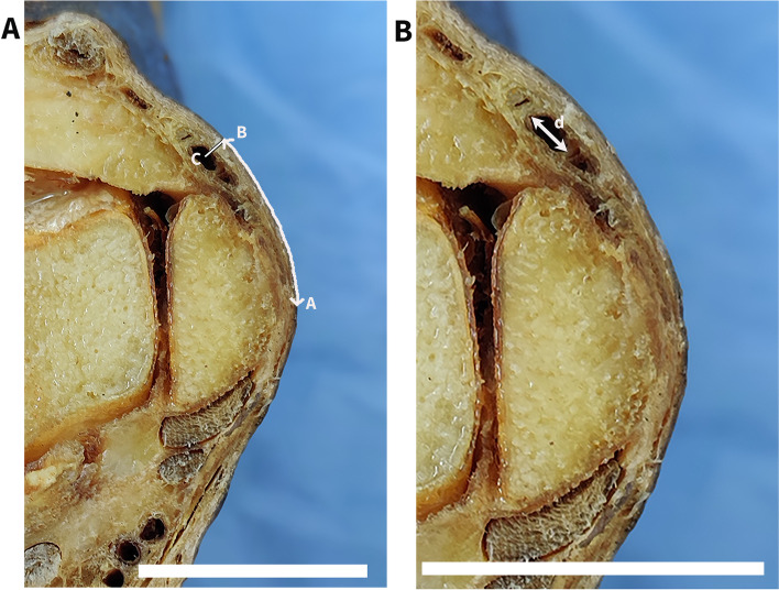
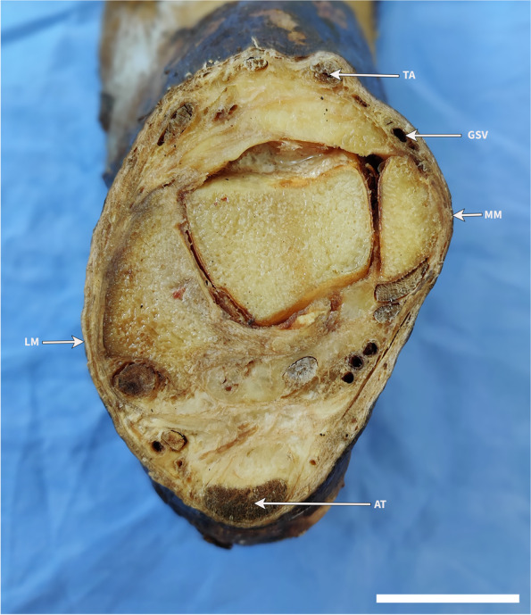
The distal great saphenous vein is a popular site for venous access by means of percutaneous cannulation or venous cutdown in a hemodynamically unstable patient. The aim of this study was to precisely define the surface anatomy and dimensions of the distal part of the great saphenous vein to facilitate the aforementioned procedures. Cross-sectional anatomy of the distal saphenous vein was studied in 24 cadaveric ankles sectioned at a horizontal plane across the most prominent points of the medial and lateral malleoli. The curvilinear distance from the most prominent point of the medial malleolus to the center of the saphenous vein, its widest collapsed diameter and skin depth were obtained. The great saphenous vein was located at a mean distance of 24.4 ± 7.9 mm anterior to the medial malleolus. The mean widest collapsed diameter was 3.8 ± 1.5 mm. The mean distance from the skin surface to the vein was 4.1 ± 1.2 mm. These measurements could be used to locate the saphenous vein accurately, particularly in hemodynamically unstable patients with visually indiscernible veins.


 分享
分享
 求助内容:
求助内容: 应助结果提醒方式:
 扫码关注我们
