{"title":"计算机断层扫描血管造影术确定的左心耳闭合最佳荧光透视投影角度的效率。","authors":"","doi":"10.1016/j.hjc.2023.09.009","DOIUrl":null,"url":null,"abstract":"<div><h3>Background</h3><p>Left atrial appendage (LAA) closure (LAAC) procedures are conventionally performed using empirical fluoroscopic viewing angles. However, because the LAA is a highly variable anatomical structure, these angles cannot depict the LAA in the optimal position. The present study aimed to assess the efficiency of using a novel optimal fluoroscopic projection angle (OPA) for LAAC and to validate its feasibility.</p></div><div><h3>Methods</h3><p>The OPAs of the derivation cohort were acquired using cardiac computed tomography angiography (CCTA) to assess its superiority for depicting LAA depth versus traditional working angles (TAs) of RAO 30°, CAU 20°. The practicability of OPA-guided LAAC was demonstrated by comparison between clinical data from the validation cohort and those from a propensity-score matched (PSM) control group, as well as randomized controlled studies investigating LAAC.</p></div><div><h3>Results</h3><p>Of 705 patients in the derivation cohort, the median OPA was RAO 46°, CAU 31°. Compared with TA, the OPA depicted a longer mean (±SD) LAA depth (5.1 ± 4.4) mm and a larger orifice diameter (1.1 ± 1.1 mm), (P < 0.0001 for both). All 38 OPA-guided LAACs were successful, with a shorter mean procedure duration (42.9 ± 12.3 min versus [vs.] 107.2 ± 41.5 min; P < 0.0001) and reduced device consumption (1.08 vs. 1.5 per case), compared with the PSM control group. At the 3-month follow-up, the incidence of peri-device leak was 52.6% (20/38) detected by CCTA, with a mean leakage of 1.6 ± 0.8 mm.</p></div><div><h3>Conclusion</h3><p>By unfolding the LAA depth and orifice diameter for a better view, OPA demonstrated the potential to optimize LAAC procedural efficiency, although further larger-scale studies are required to confirm this.</p></div>","PeriodicalId":55062,"journal":{"name":"Hellenic Journal of Cardiology","volume":"78 ","pages":"Pages 50-59"},"PeriodicalIF":3.0000,"publicationDate":"2024-07-01","publicationTypes":"Journal Article","fieldsOfStudy":null,"isOpenAccess":false,"openAccessPdf":"https://www.sciencedirect.com/science/article/pii/S1109966623001793/pdfft?md5=86a89c29f25632e2842899cd3a3c3626&pid=1-s2.0-S1109966623001793-main.pdf","citationCount":"0","resultStr":"{\"title\":\"Efficiency of optimal fluoroscopic projection angle defined by computed tomography angiography for left atrial appendage closure\",\"authors\":\"\",\"doi\":\"10.1016/j.hjc.2023.09.009\",\"DOIUrl\":null,\"url\":null,\"abstract\":\"<div><h3>Background</h3><p>Left atrial appendage (LAA) closure (LAAC) procedures are conventionally performed using empirical fluoroscopic viewing angles. However, because the LAA is a highly variable anatomical structure, these angles cannot depict the LAA in the optimal position. The present study aimed to assess the efficiency of using a novel optimal fluoroscopic projection angle (OPA) for LAAC and to validate its feasibility.</p></div><div><h3>Methods</h3><p>The OPAs of the derivation cohort were acquired using cardiac computed tomography angiography (CCTA) to assess its superiority for depicting LAA depth versus traditional working angles (TAs) of RAO 30°, CAU 20°. The practicability of OPA-guided LAAC was demonstrated by comparison between clinical data from the validation cohort and those from a propensity-score matched (PSM) control group, as well as randomized controlled studies investigating LAAC.</p></div><div><h3>Results</h3><p>Of 705 patients in the derivation cohort, the median OPA was RAO 46°, CAU 31°. Compared with TA, the OPA depicted a longer mean (±SD) LAA depth (5.1 ± 4.4) mm and a larger orifice diameter (1.1 ± 1.1 mm), (P < 0.0001 for both). All 38 OPA-guided LAACs were successful, with a shorter mean procedure duration (42.9 ± 12.3 min versus [vs.] 107.2 ± 41.5 min; P < 0.0001) and reduced device consumption (1.08 vs. 1.5 per case), compared with the PSM control group. At the 3-month follow-up, the incidence of peri-device leak was 52.6% (20/38) detected by CCTA, with a mean leakage of 1.6 ± 0.8 mm.</p></div><div><h3>Conclusion</h3><p>By unfolding the LAA depth and orifice diameter for a better view, OPA demonstrated the potential to optimize LAAC procedural efficiency, although further larger-scale studies are required to confirm this.</p></div>\",\"PeriodicalId\":55062,\"journal\":{\"name\":\"Hellenic Journal of Cardiology\",\"volume\":\"78 \",\"pages\":\"Pages 50-59\"},\"PeriodicalIF\":3.0000,\"publicationDate\":\"2024-07-01\",\"publicationTypes\":\"Journal Article\",\"fieldsOfStudy\":null,\"isOpenAccess\":false,\"openAccessPdf\":\"https://www.sciencedirect.com/science/article/pii/S1109966623001793/pdfft?md5=86a89c29f25632e2842899cd3a3c3626&pid=1-s2.0-S1109966623001793-main.pdf\",\"citationCount\":\"0\",\"resultStr\":null,\"platform\":\"Semanticscholar\",\"paperid\":null,\"PeriodicalName\":\"Hellenic Journal of Cardiology\",\"FirstCategoryId\":\"3\",\"ListUrlMain\":\"https://www.sciencedirect.com/science/article/pii/S1109966623001793\",\"RegionNum\":3,\"RegionCategory\":\"医学\",\"ArticlePicture\":[],\"TitleCN\":null,\"AbstractTextCN\":null,\"PMCID\":null,\"EPubDate\":\"2023/9/15 0:00:00\",\"PubModel\":\"Epub\",\"JCR\":\"Q2\",\"JCRName\":\"CARDIAC & CARDIOVASCULAR SYSTEMS\",\"Score\":null,\"Total\":0}","platform":"Semanticscholar","paperid":null,"PeriodicalName":"Hellenic Journal of Cardiology","FirstCategoryId":"3","ListUrlMain":"https://www.sciencedirect.com/science/article/pii/S1109966623001793","RegionNum":3,"RegionCategory":"医学","ArticlePicture":[],"TitleCN":null,"AbstractTextCN":null,"PMCID":null,"EPubDate":"2023/9/15 0:00:00","PubModel":"Epub","JCR":"Q2","JCRName":"CARDIAC & CARDIOVASCULAR SYSTEMS","Score":null,"Total":0}
Efficiency of optimal fluoroscopic projection angle defined by computed tomography angiography for left atrial appendage closure
Background
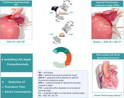
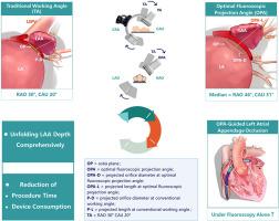
Left atrial appendage (LAA) closure (LAAC) procedures are conventionally performed using empirical fluoroscopic viewing angles. However, because the LAA is a highly variable anatomical structure, these angles cannot depict the LAA in the optimal position. The present study aimed to assess the efficiency of using a novel optimal fluoroscopic projection angle (OPA) for LAAC and to validate its feasibility.
Methods
The OPAs of the derivation cohort were acquired using cardiac computed tomography angiography (CCTA) to assess its superiority for depicting LAA depth versus traditional working angles (TAs) of RAO 30°, CAU 20°. The practicability of OPA-guided LAAC was demonstrated by comparison between clinical data from the validation cohort and those from a propensity-score matched (PSM) control group, as well as randomized controlled studies investigating LAAC.
Results
Of 705 patients in the derivation cohort, the median OPA was RAO 46°, CAU 31°. Compared with TA, the OPA depicted a longer mean (±SD) LAA depth (5.1 ± 4.4) mm and a larger orifice diameter (1.1 ± 1.1 mm), (P < 0.0001 for both). All 38 OPA-guided LAACs were successful, with a shorter mean procedure duration (42.9 ± 12.3 min versus [vs.] 107.2 ± 41.5 min; P < 0.0001) and reduced device consumption (1.08 vs. 1.5 per case), compared with the PSM control group. At the 3-month follow-up, the incidence of peri-device leak was 52.6% (20/38) detected by CCTA, with a mean leakage of 1.6 ± 0.8 mm.
Conclusion
By unfolding the LAA depth and orifice diameter for a better view, OPA demonstrated the potential to optimize LAAC procedural efficiency, although further larger-scale studies are required to confirm this.
期刊介绍:
The Hellenic Journal of Cardiology (International Edition, ISSN 1109-9666) is the official journal of the Hellenic Society of Cardiology and aims to publish high-quality articles on all aspects of cardiovascular medicine. A primary goal is to publish in each issue a number of original articles related to clinical and basic research. Many of these will be accompanied by invited editorial comments.
Hot topics, such as molecular cardiology, and innovative cardiac imaging and electrophysiological mapping techniques, will appear frequently in the journal in the form of invited expert articles or special reports. The Editorial Committee also attaches great importance to subjects related to continuing medical education, the implementation of guidelines and cost effectiveness in cardiology.


 分享
分享
 求助内容:
求助内容: 应助结果提醒方式:
 扫码关注我们
