Si-Si Qi, Ying Miao, You-Yu Sheng, Rui-Ming Hu, Jun Zhao, Qin-Ping Yang
{"title":"MicroRNA-1246抑制NFATc1磷酸化并调节T辅助17细胞活化在重度斑秃发病中的作用","authors":"Si-Si Qi, Ying Miao, You-Yu Sheng, Rui-Ming Hu, Jun Zhao, Qin-Ping Yang","doi":"10.5021/ad.22.126","DOIUrl":null,"url":null,"abstract":"<p><strong>Background: </strong>We found microRNA (miR)-1246 to be significantly differentially expressed between severe active alopecia areata (AA) patients and healthy individuals.</p><p><strong>Objective: </strong>To explore the role and mechanism of miR-1246 in severe AA.</p><p><strong>Methods: </strong>Expression of miR-1246, dual-specific tyrosine phosphorylation-regulated kinase 1A (DYRK1A), and nuclear factor of activated T cells 1c (NFATc1) in peripheral CD4<sup>+</sup> T cells and in scalp tissues of patients were detected using RT-qPCR, Western blot, and immunohistochemistry assays. Peripheral CD4<sup>+</sup> T cells from the AA patients were transfected with lentiviral vectors overexpressing miR-1246. RT-qPCR and Western blot analysis were used to measure mRNA or protein expression of retinoic-acid-receptor-related orphan nuclear receptor gamma (ROR-γt), interleukin (IL)-17, DYRK1A, NFATc1, and phosphorylated NFATc1. Flow cytometry was used to assay the CD4<sup>+</sup>IL-17<sup>+</sup> cells proportion. ELISA was used to measure cytokine levels.</p><p><strong>Results: </strong>miR-1246 levels decreased and DYRK1A and NFATc1 mRNA levels significantly increased in the peripheral CD4<sup>+</sup> T cells and scalp tissues of severe active AA samples. NFATc1 protein expression was also significantly increased in the peripheral CD4<sup>+</sup> T cells but not in the scalp tissues. NFATc1 positive cells were mainly distributed among infiltrating inflammatory cells around hair follicles. In peripheral CD4<sup>+</sup> T cells of severe active AA, overexpression of miR-1246 resulted in significant downregulation of DYRK1A, NFATc1, ROR-γt, and IL-17 mRNA and phosphorylated NFATc1 protein, as well as a decrease in the CD4<sup>+</sup>IL-17<sup>+</sup> cells proportion and the IL-17F level.</p><p><strong>Conclusion: </strong>miR-1246 can inhibit NFAT signaling and Th17 cell activation, which may be beneficial in the severe AA treatment.</p>","PeriodicalId":8233,"journal":{"name":"Annals of Dermatology","volume":"35 1","pages":"46-55"},"PeriodicalIF":1.3000,"publicationDate":"2023-02-01","publicationTypes":"Journal Article","fieldsOfStudy":null,"isOpenAccess":false,"openAccessPdf":"https://ftp.ncbi.nlm.nih.gov/pub/pmc/oa_pdf/62/94/ad-35-46.PMC9905862.pdf","citationCount":"0","resultStr":"{\"title\":\"MicroRNA-1246 Inhibits NFATc1 Phosphorylation and Regulates T Helper 17 Cell Activation in the Pathogenesis of Severe Alopecia Areata.\",\"authors\":\"Si-Si Qi, Ying Miao, You-Yu Sheng, Rui-Ming Hu, Jun Zhao, Qin-Ping Yang\",\"doi\":\"10.5021/ad.22.126\",\"DOIUrl\":null,\"url\":null,\"abstract\":\"<p><strong>Background: </strong>We found microRNA (miR)-1246 to be significantly differentially expressed between severe active alopecia areata (AA) patients and healthy individuals.</p><p><strong>Objective: </strong>To explore the role and mechanism of miR-1246 in severe AA.</p><p><strong>Methods: </strong>Expression of miR-1246, dual-specific tyrosine phosphorylation-regulated kinase 1A (DYRK1A), and nuclear factor of activated T cells 1c (NFATc1) in peripheral CD4<sup>+</sup> T cells and in scalp tissues of patients were detected using RT-qPCR, Western blot, and immunohistochemistry assays. Peripheral CD4<sup>+</sup> T cells from the AA patients were transfected with lentiviral vectors overexpressing miR-1246. RT-qPCR and Western blot analysis were used to measure mRNA or protein expression of retinoic-acid-receptor-related orphan nuclear receptor gamma (ROR-γt), interleukin (IL)-17, DYRK1A, NFATc1, and phosphorylated NFATc1. Flow cytometry was used to assay the CD4<sup>+</sup>IL-17<sup>+</sup> cells proportion. ELISA was used to measure cytokine levels.</p><p><strong>Results: </strong>miR-1246 levels decreased and DYRK1A and NFATc1 mRNA levels significantly increased in the peripheral CD4<sup>+</sup> T cells and scalp tissues of severe active AA samples. NFATc1 protein expression was also significantly increased in the peripheral CD4<sup>+</sup> T cells but not in the scalp tissues. NFATc1 positive cells were mainly distributed among infiltrating inflammatory cells around hair follicles. In peripheral CD4<sup>+</sup> T cells of severe active AA, overexpression of miR-1246 resulted in significant downregulation of DYRK1A, NFATc1, ROR-γt, and IL-17 mRNA and phosphorylated NFATc1 protein, as well as a decrease in the CD4<sup>+</sup>IL-17<sup>+</sup> cells proportion and the IL-17F level.</p><p><strong>Conclusion: </strong>miR-1246 can inhibit NFAT signaling and Th17 cell activation, which may be beneficial in the severe AA treatment.</p>\",\"PeriodicalId\":8233,\"journal\":{\"name\":\"Annals of Dermatology\",\"volume\":\"35 1\",\"pages\":\"46-55\"},\"PeriodicalIF\":1.3000,\"publicationDate\":\"2023-02-01\",\"publicationTypes\":\"Journal Article\",\"fieldsOfStudy\":null,\"isOpenAccess\":false,\"openAccessPdf\":\"https://ftp.ncbi.nlm.nih.gov/pub/pmc/oa_pdf/62/94/ad-35-46.PMC9905862.pdf\",\"citationCount\":\"0\",\"resultStr\":null,\"platform\":\"Semanticscholar\",\"paperid\":null,\"PeriodicalName\":\"Annals of Dermatology\",\"FirstCategoryId\":\"3\",\"ListUrlMain\":\"https://doi.org/10.5021/ad.22.126\",\"RegionNum\":4,\"RegionCategory\":\"医学\",\"ArticlePicture\":[],\"TitleCN\":null,\"AbstractTextCN\":null,\"PMCID\":null,\"EPubDate\":\"\",\"PubModel\":\"\",\"JCR\":\"Q3\",\"JCRName\":\"DERMATOLOGY\",\"Score\":null,\"Total\":0}","platform":"Semanticscholar","paperid":null,"PeriodicalName":"Annals of Dermatology","FirstCategoryId":"3","ListUrlMain":"https://doi.org/10.5021/ad.22.126","RegionNum":4,"RegionCategory":"医学","ArticlePicture":[],"TitleCN":null,"AbstractTextCN":null,"PMCID":null,"EPubDate":"","PubModel":"","JCR":"Q3","JCRName":"DERMATOLOGY","Score":null,"Total":0}
MicroRNA-1246 Inhibits NFATc1 Phosphorylation and Regulates T Helper 17 Cell Activation in the Pathogenesis of Severe Alopecia Areata.
Background: We found microRNA (miR)-1246 to be significantly differentially expressed between severe active alopecia areata (AA) patients and healthy individuals.
Objective: To explore the role and mechanism of miR-1246 in severe AA.
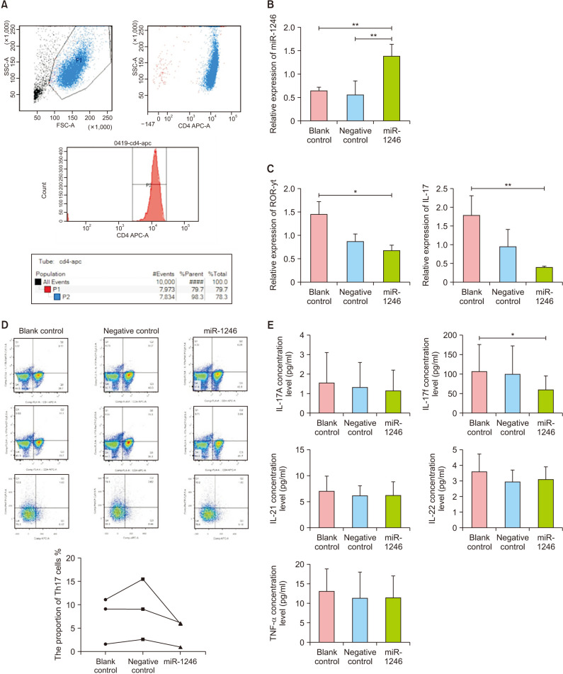
Methods: Expression of miR-1246, dual-specific tyrosine phosphorylation-regulated kinase 1A (DYRK1A), and nuclear factor of activated T cells 1c (NFATc1) in peripheral CD4+ T cells and in scalp tissues of patients were detected using RT-qPCR, Western blot, and immunohistochemistry assays. Peripheral CD4+ T cells from the AA patients were transfected with lentiviral vectors overexpressing miR-1246. RT-qPCR and Western blot analysis were used to measure mRNA or protein expression of retinoic-acid-receptor-related orphan nuclear receptor gamma (ROR-γt), interleukin (IL)-17, DYRK1A, NFATc1, and phosphorylated NFATc1. Flow cytometry was used to assay the CD4+IL-17+ cells proportion. ELISA was used to measure cytokine levels.
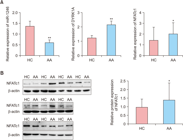
Results: miR-1246 levels decreased and DYRK1A and NFATc1 mRNA levels significantly increased in the peripheral CD4+ T cells and scalp tissues of severe active AA samples. NFATc1 protein expression was also significantly increased in the peripheral CD4+ T cells but not in the scalp tissues. NFATc1 positive cells were mainly distributed among infiltrating inflammatory cells around hair follicles. In peripheral CD4+ T cells of severe active AA, overexpression of miR-1246 resulted in significant downregulation of DYRK1A, NFATc1, ROR-γt, and IL-17 mRNA and phosphorylated NFATc1 protein, as well as a decrease in the CD4+IL-17+ cells proportion and the IL-17F level.
Conclusion: miR-1246 can inhibit NFAT signaling and Th17 cell activation, which may be beneficial in the severe AA treatment.
期刊介绍:
Annals of Dermatology (Ann Dermatol) is the official peer-reviewed publication of the Korean Dermatological Association and the Korean Society for Investigative Dermatology. Since 1989, Ann Dermatol has contributed as a platform for communicating the latest research outcome and recent trend of dermatology in Korea and all over the world.
Ann Dermatol seeks for ameliorated understanding of skin and skin-related disease for clinicians and researchers. Ann Dermatol deals with diverse skin-related topics from laboratory investigations to clinical outcomes and invites review articles, original articles, case reports, brief reports and items of correspondence. Ann Dermatol is interested in contributions from all countries in which good and advanced research is carried out. Ann Dermatol willingly recruits well-organized and significant manuscripts with proper scope throughout the world.


 分享
分享
 求助内容:
求助内容: 应助结果提醒方式:
 扫码关注我们
