Prevalence and morphology of different root canal systems in mandibular premolars: a cross-sectional observational study
Background
This study investigated the prevalence and morphology of C-shaped and non-C-shaped root canal systems in permanent mandibular first (PM1) and second (PM2) premolars using retrospective analysis of cone-beam computed tomography (CBCT) scans, and panoramic radiographs.
Methods
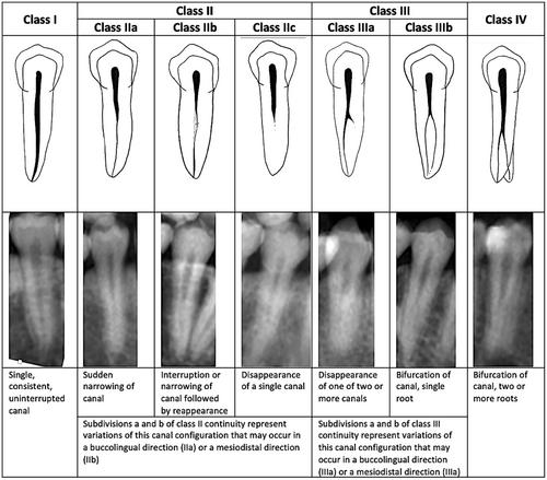
CBCT scans from 2000 patients were screened for the presence of premolars with C-shaped canals and then assessed at three axial levels to determine the canal classification. The teeth were also assessed for Vertucci configuration, number of roots, and radicular grooves. Pre-existing panoramic radiographs were evaluated to identify features specific to PM1/2 with multiple canals or C-shaped anatomy.
Results
A total of 1576 PM1 and 1424 PM2 from 880 patients were evaluated. The overall prevalence of C-shaped canals was 2.2% (3.3% PM1, 1.0% PM2), with 49 (5.6%) patients presenting with at least one C-shaped mandibular premolar. There were 2.3 ± 0.6 and 2.4 ± 0.5 different classifiable cross-sections per tooth for PM1 and PM2, respectively. The sudden disappearance or bifurcation of a canal on panoramic radiographs was associated with the presence of multiple canals (P < 0.001) or C-shaped anatomy (P = 0.03).
期刊介绍:
The Australian Dental Journal provides a forum for the exchange of information about new and significant research in dentistry, promoting the discipline of dentistry in Australia and throughout the world. It comprises peer-reviewed research articles as its core material, supplemented by reviews, theoretical articles, special features and commentaries.


 分享
分享
 求助内容:
求助内容: 应助结果提醒方式:
 扫码关注我们
