Viktor V. Nikolaev, Yury V. Kistenev, Marius Kröger, Hala Zuhayri, Maxim E. Darvin
{"title":"皮肤成纤维细胞非侵入性成像的光学方法综述——从体外到离体和体内可视化。","authors":"Viktor V. Nikolaev, Yury V. Kistenev, Marius Kröger, Hala Zuhayri, Maxim E. Darvin","doi":"10.1002/jbio.202300223","DOIUrl":null,"url":null,"abstract":"<p>Fibroblasts are among the most common cell types in the stroma responsible for creating and maintaining the structural organization of the extracellular matrix in the dermis, skin regeneration, and a range of immune responses. Until now, the processes of fibroblast adaptation and functioning in a varying environment have not been fully understood. Modern laser microscopes are capable of studying fibroblasts in vitro and ex vivo. One-photon- and two-photon-excited fluorescence microscopy, Raman spectroscopy/microspectroscopy are well-suited noninvasive optical methods for fibroblast imaging in vitro and ex vivo. In vivo staining-free fibroblast imaging is not still implemented. The exception is fibroblast imaging in tattooed skin. Although in vivo noninvasive staining-free imaging of fibroblasts in the skin has not yet been implemented, it is expected in the future. This review summarizes the state-of-the-art in fibroblast visualization using optical methods and discusses the advantages, limitations, and prospects for future noninvasive imaging.</p>","PeriodicalId":184,"journal":{"name":"Journal of Biophotonics","volume":"17 1","pages":""},"PeriodicalIF":2.0000,"publicationDate":"2023-11-29","publicationTypes":"Journal Article","fieldsOfStudy":null,"isOpenAccess":false,"openAccessPdf":"","citationCount":"0","resultStr":"{\"title\":\"Review of optical methods for noninvasive imaging of skin fibroblasts—From in vitro to ex vivo and in vivo visualization\",\"authors\":\"Viktor V. Nikolaev, Yury V. Kistenev, Marius Kröger, Hala Zuhayri, Maxim E. Darvin\",\"doi\":\"10.1002/jbio.202300223\",\"DOIUrl\":null,\"url\":null,\"abstract\":\"<p>Fibroblasts are among the most common cell types in the stroma responsible for creating and maintaining the structural organization of the extracellular matrix in the dermis, skin regeneration, and a range of immune responses. Until now, the processes of fibroblast adaptation and functioning in a varying environment have not been fully understood. Modern laser microscopes are capable of studying fibroblasts in vitro and ex vivo. One-photon- and two-photon-excited fluorescence microscopy, Raman spectroscopy/microspectroscopy are well-suited noninvasive optical methods for fibroblast imaging in vitro and ex vivo. In vivo staining-free fibroblast imaging is not still implemented. The exception is fibroblast imaging in tattooed skin. Although in vivo noninvasive staining-free imaging of fibroblasts in the skin has not yet been implemented, it is expected in the future. This review summarizes the state-of-the-art in fibroblast visualization using optical methods and discusses the advantages, limitations, and prospects for future noninvasive imaging.</p>\",\"PeriodicalId\":184,\"journal\":{\"name\":\"Journal of Biophotonics\",\"volume\":\"17 1\",\"pages\":\"\"},\"PeriodicalIF\":2.0000,\"publicationDate\":\"2023-11-29\",\"publicationTypes\":\"Journal Article\",\"fieldsOfStudy\":null,\"isOpenAccess\":false,\"openAccessPdf\":\"\",\"citationCount\":\"0\",\"resultStr\":null,\"platform\":\"Semanticscholar\",\"paperid\":null,\"PeriodicalName\":\"Journal of Biophotonics\",\"FirstCategoryId\":\"101\",\"ListUrlMain\":\"https://onlinelibrary.wiley.com/doi/10.1002/jbio.202300223\",\"RegionNum\":3,\"RegionCategory\":\"物理与天体物理\",\"ArticlePicture\":[],\"TitleCN\":null,\"AbstractTextCN\":null,\"PMCID\":null,\"EPubDate\":\"\",\"PubModel\":\"\",\"JCR\":\"Q3\",\"JCRName\":\"BIOCHEMICAL RESEARCH METHODS\",\"Score\":null,\"Total\":0}","platform":"Semanticscholar","paperid":null,"PeriodicalName":"Journal of Biophotonics","FirstCategoryId":"101","ListUrlMain":"https://onlinelibrary.wiley.com/doi/10.1002/jbio.202300223","RegionNum":3,"RegionCategory":"物理与天体物理","ArticlePicture":[],"TitleCN":null,"AbstractTextCN":null,"PMCID":null,"EPubDate":"","PubModel":"","JCR":"Q3","JCRName":"BIOCHEMICAL RESEARCH METHODS","Score":null,"Total":0}
Review of optical methods for noninvasive imaging of skin fibroblasts—From in vitro to ex vivo and in vivo visualization
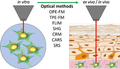
Fibroblasts are among the most common cell types in the stroma responsible for creating and maintaining the structural organization of the extracellular matrix in the dermis, skin regeneration, and a range of immune responses. Until now, the processes of fibroblast adaptation and functioning in a varying environment have not been fully understood. Modern laser microscopes are capable of studying fibroblasts in vitro and ex vivo. One-photon- and two-photon-excited fluorescence microscopy, Raman spectroscopy/microspectroscopy are well-suited noninvasive optical methods for fibroblast imaging in vitro and ex vivo. In vivo staining-free fibroblast imaging is not still implemented. The exception is fibroblast imaging in tattooed skin. Although in vivo noninvasive staining-free imaging of fibroblasts in the skin has not yet been implemented, it is expected in the future. This review summarizes the state-of-the-art in fibroblast visualization using optical methods and discusses the advantages, limitations, and prospects for future noninvasive imaging.
期刊介绍:
The first international journal dedicated to publishing reviews and original articles from this exciting field, the Journal of Biophotonics covers the broad range of research on interactions between light and biological material. The journal offers a platform where the physicist communicates with the biologist and where the clinical practitioner learns about the latest tools for the diagnosis of diseases. As such, the journal is highly interdisciplinary, publishing cutting edge research in the fields of life sciences, medicine, physics, chemistry, and engineering. The coverage extends from fundamental research to specific developments, while also including the latest applications.


 分享
分享
 求助内容:
求助内容: 应助结果提醒方式:
 扫码关注我们
