Yunke Ren, Xinying Chu, Janaka Senarathna, Akanksha Bhargava, Warren L. Grayson, Arvind P. Pathak
{"title":"多模态成像显示颅骨骨缺损愈合过程中血管生成的体内进化。","authors":"Yunke Ren, Xinying Chu, Janaka Senarathna, Akanksha Bhargava, Warren L. Grayson, Arvind P. Pathak","doi":"10.1007/s10456-023-09899-0","DOIUrl":null,"url":null,"abstract":"<div><p>The healing of calvarial bone defects is a pressing clinical problem that involves the dynamic interplay between angiogenesis and osteogenesis within the osteogenic niche. Although structural and functional vascular remodeling (i.e., angiogenic evolution) in the osteogenic niche is a crucial modulator of oxygenation, inflammatory and bone precursor cells, most clinical and pre-clinical investigations have been limited to characterizing structural changes in the vasculature and bone. Therefore, we developed a new multimodality imaging approach that for the first time enabled the longitudinal (i.e., over four weeks) and dynamic characterization of multiple in vivo functional parameters in the remodeled vasculature and its effects on de novo osteogenesis, in a preclinical calvarial defect model. We employed multi-wavelength intrinsic optical signal (IOS) imaging to assess microvascular remodeling, intravascular oxygenation (SO<sub>2</sub>), and osteogenesis; laser speckle contrast (LSC) imaging to assess concomitant changes in blood flow and vascular maturity; and micro-computed tomography (μCT) to validate volumetric changes in calvarial bone. We found that angiogenic evolution was tightly coupled with calvarial bone regeneration and corresponded to distinct phases of bone healing, such as injury, hematoma formation, revascularization, and remodeling. The first three phases occurred during the initial two weeks of bone healing and were characterized by significant in vivo changes in vascular morphology, blood flow, oxygenation, and maturity. Overall, angiogenic evolution preceded osteogenesis, which only plateaued toward the end of bone healing (i.e., four weeks). Collectively, these data indicate the crucial role of angiogenic evolution in osteogenesis. We believe that such multimodality imaging approaches have the potential to inform the design of more efficacious tissue-engineering calvarial defect treatments.</p></div>","PeriodicalId":7886,"journal":{"name":"Angiogenesis","volume":"27 1","pages":"105 - 119"},"PeriodicalIF":9.2000,"publicationDate":"2023-11-30","publicationTypes":"Journal Article","fieldsOfStudy":null,"isOpenAccess":false,"openAccessPdf":"","citationCount":"0","resultStr":"{\"title\":\"Multimodality imaging reveals angiogenic evolution in vivo during calvarial bone defect healing\",\"authors\":\"Yunke Ren, Xinying Chu, Janaka Senarathna, Akanksha Bhargava, Warren L. Grayson, Arvind P. Pathak\",\"doi\":\"10.1007/s10456-023-09899-0\",\"DOIUrl\":null,\"url\":null,\"abstract\":\"<div><p>The healing of calvarial bone defects is a pressing clinical problem that involves the dynamic interplay between angiogenesis and osteogenesis within the osteogenic niche. Although structural and functional vascular remodeling (i.e., angiogenic evolution) in the osteogenic niche is a crucial modulator of oxygenation, inflammatory and bone precursor cells, most clinical and pre-clinical investigations have been limited to characterizing structural changes in the vasculature and bone. Therefore, we developed a new multimodality imaging approach that for the first time enabled the longitudinal (i.e., over four weeks) and dynamic characterization of multiple in vivo functional parameters in the remodeled vasculature and its effects on de novo osteogenesis, in a preclinical calvarial defect model. We employed multi-wavelength intrinsic optical signal (IOS) imaging to assess microvascular remodeling, intravascular oxygenation (SO<sub>2</sub>), and osteogenesis; laser speckle contrast (LSC) imaging to assess concomitant changes in blood flow and vascular maturity; and micro-computed tomography (μCT) to validate volumetric changes in calvarial bone. We found that angiogenic evolution was tightly coupled with calvarial bone regeneration and corresponded to distinct phases of bone healing, such as injury, hematoma formation, revascularization, and remodeling. The first three phases occurred during the initial two weeks of bone healing and were characterized by significant in vivo changes in vascular morphology, blood flow, oxygenation, and maturity. Overall, angiogenic evolution preceded osteogenesis, which only plateaued toward the end of bone healing (i.e., four weeks). Collectively, these data indicate the crucial role of angiogenic evolution in osteogenesis. We believe that such multimodality imaging approaches have the potential to inform the design of more efficacious tissue-engineering calvarial defect treatments.</p></div>\",\"PeriodicalId\":7886,\"journal\":{\"name\":\"Angiogenesis\",\"volume\":\"27 1\",\"pages\":\"105 - 119\"},\"PeriodicalIF\":9.2000,\"publicationDate\":\"2023-11-30\",\"publicationTypes\":\"Journal Article\",\"fieldsOfStudy\":null,\"isOpenAccess\":false,\"openAccessPdf\":\"\",\"citationCount\":\"0\",\"resultStr\":null,\"platform\":\"Semanticscholar\",\"paperid\":null,\"PeriodicalName\":\"Angiogenesis\",\"FirstCategoryId\":\"3\",\"ListUrlMain\":\"https://link.springer.com/article/10.1007/s10456-023-09899-0\",\"RegionNum\":1,\"RegionCategory\":\"医学\",\"ArticlePicture\":[],\"TitleCN\":null,\"AbstractTextCN\":null,\"PMCID\":null,\"EPubDate\":\"\",\"PubModel\":\"\",\"JCR\":\"Q1\",\"JCRName\":\"PERIPHERAL VASCULAR DISEASE\",\"Score\":null,\"Total\":0}","platform":"Semanticscholar","paperid":null,"PeriodicalName":"Angiogenesis","FirstCategoryId":"3","ListUrlMain":"https://link.springer.com/article/10.1007/s10456-023-09899-0","RegionNum":1,"RegionCategory":"医学","ArticlePicture":[],"TitleCN":null,"AbstractTextCN":null,"PMCID":null,"EPubDate":"","PubModel":"","JCR":"Q1","JCRName":"PERIPHERAL VASCULAR DISEASE","Score":null,"Total":0}
Multimodality imaging reveals angiogenic evolution in vivo during calvarial bone defect healing
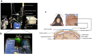
The healing of calvarial bone defects is a pressing clinical problem that involves the dynamic interplay between angiogenesis and osteogenesis within the osteogenic niche. Although structural and functional vascular remodeling (i.e., angiogenic evolution) in the osteogenic niche is a crucial modulator of oxygenation, inflammatory and bone precursor cells, most clinical and pre-clinical investigations have been limited to characterizing structural changes in the vasculature and bone. Therefore, we developed a new multimodality imaging approach that for the first time enabled the longitudinal (i.e., over four weeks) and dynamic characterization of multiple in vivo functional parameters in the remodeled vasculature and its effects on de novo osteogenesis, in a preclinical calvarial defect model. We employed multi-wavelength intrinsic optical signal (IOS) imaging to assess microvascular remodeling, intravascular oxygenation (SO2), and osteogenesis; laser speckle contrast (LSC) imaging to assess concomitant changes in blood flow and vascular maturity; and micro-computed tomography (μCT) to validate volumetric changes in calvarial bone. We found that angiogenic evolution was tightly coupled with calvarial bone regeneration and corresponded to distinct phases of bone healing, such as injury, hematoma formation, revascularization, and remodeling. The first three phases occurred during the initial two weeks of bone healing and were characterized by significant in vivo changes in vascular morphology, blood flow, oxygenation, and maturity. Overall, angiogenic evolution preceded osteogenesis, which only plateaued toward the end of bone healing (i.e., four weeks). Collectively, these data indicate the crucial role of angiogenic evolution in osteogenesis. We believe that such multimodality imaging approaches have the potential to inform the design of more efficacious tissue-engineering calvarial defect treatments.
期刊介绍:
Angiogenesis, a renowned international journal, seeks to publish high-quality original articles and reviews on the cellular and molecular mechanisms governing angiogenesis in both normal and pathological conditions. By serving as a primary platform for swift communication within the field of angiogenesis research, this multidisciplinary journal showcases pioneering experimental studies utilizing molecular techniques, in vitro methods, animal models, and clinical investigations into angiogenic diseases. Furthermore, Angiogenesis sheds light on cutting-edge therapeutic strategies for promoting or inhibiting angiogenesis, while also highlighting fresh markers and techniques for disease diagnosis and prognosis.


 分享
分享
 求助内容:
求助内容: 应助结果提醒方式:
 扫码关注我们
