Stephan Dreschers, Christopher Platen, Louise Oppermann, Caitlin Doughty, Andreas Ludwig, Aaron Babendreyer, Thorsten W Orlikowsky
{"title":"抗双调节蛋白egf受体(AREG)影响单核细胞和T细胞的共刺激分子并调节T细胞反应。","authors":"Stephan Dreschers, Christopher Platen, Louise Oppermann, Caitlin Doughty, Andreas Ludwig, Aaron Babendreyer, Thorsten W Orlikowsky","doi":"10.1155/2023/8883045","DOIUrl":null,"url":null,"abstract":"<p><p>Amphiregulin (AREG) is a ligand of the epidermal growth factor receptor (EGFR) and has been shown to regulate the phagocytosis-induced cell death of monocytes in peripheral blood. AREG-dependent apoptotic signaling engages factors of the intrinsic and extrinsic apoptotic pathway, such as BCL-2, BCL-XL, and death ligand/receptor CD95/CD95L. Here, we tested the hypothesis that AREG influences costimulatory monocyte functions, which are crucial for T-cell responses. We found a stronger expression of AREG and EGFR in monocytes compared to lymphocytes. As a novel function of AREG, we observed reduced T-cell proliferation following polyclonal T-cell stimulation with OKT3. This reduction of proliferation occurred in the presence of monocytes as well as in their absence, monocyte signaling being replaced by crosslinking of OKT3. Increasing concentrations of AREG down-modulated the concentration of costimulatory B7 molecules (CD80/CD86) and HLA-DR on monocytes. In proliferation assays, CD28 expression on T cells was down-modulated on the application of OKT3 but unaltered by AREG. LcK activation, following OKT3-stimulation, was reduced in T cells that had been coincubated with AREG. The effects of AREG on T-cell phenotypes were also present when monocytes were depleted and OKT3 was crosslinked. The rearranged expression of immunological synapse proteins was accompanied by an alteration of T-cell polarization. Although the proportion of regulatory T cells was not shifted by AREG, IL-17-expressing T cells were significantly enhanced, with a bias toward TH1-polarization. Taken together, these results suggest that AREG acts as an immunoregulatory molecule at the interface between antigen-presenting cells and T cells.</p>","PeriodicalId":15952,"journal":{"name":"Journal of Immunology Research","volume":"2023 ","pages":"8883045"},"PeriodicalIF":3.6000,"publicationDate":"2023-11-24","publicationTypes":"Journal Article","fieldsOfStudy":null,"isOpenAccess":false,"openAccessPdf":"https://www.ncbi.nlm.nih.gov/pmc/articles/PMC10691888/pdf/","citationCount":"0","resultStr":"{\"title\":\"EGF-Receptor against Amphiregulin (AREG) Influences Costimulatory Molecules on Monocytes and T Cells and Modulates T-Cell Responses.\",\"authors\":\"Stephan Dreschers, Christopher Platen, Louise Oppermann, Caitlin Doughty, Andreas Ludwig, Aaron Babendreyer, Thorsten W Orlikowsky\",\"doi\":\"10.1155/2023/8883045\",\"DOIUrl\":null,\"url\":null,\"abstract\":\"<p><p>Amphiregulin (AREG) is a ligand of the epidermal growth factor receptor (EGFR) and has been shown to regulate the phagocytosis-induced cell death of monocytes in peripheral blood. AREG-dependent apoptotic signaling engages factors of the intrinsic and extrinsic apoptotic pathway, such as BCL-2, BCL-XL, and death ligand/receptor CD95/CD95L. Here, we tested the hypothesis that AREG influences costimulatory monocyte functions, which are crucial for T-cell responses. We found a stronger expression of AREG and EGFR in monocytes compared to lymphocytes. As a novel function of AREG, we observed reduced T-cell proliferation following polyclonal T-cell stimulation with OKT3. This reduction of proliferation occurred in the presence of monocytes as well as in their absence, monocyte signaling being replaced by crosslinking of OKT3. Increasing concentrations of AREG down-modulated the concentration of costimulatory B7 molecules (CD80/CD86) and HLA-DR on monocytes. In proliferation assays, CD28 expression on T cells was down-modulated on the application of OKT3 but unaltered by AREG. LcK activation, following OKT3-stimulation, was reduced in T cells that had been coincubated with AREG. The effects of AREG on T-cell phenotypes were also present when monocytes were depleted and OKT3 was crosslinked. The rearranged expression of immunological synapse proteins was accompanied by an alteration of T-cell polarization. Although the proportion of regulatory T cells was not shifted by AREG, IL-17-expressing T cells were significantly enhanced, with a bias toward TH1-polarization. Taken together, these results suggest that AREG acts as an immunoregulatory molecule at the interface between antigen-presenting cells and T cells.</p>\",\"PeriodicalId\":15952,\"journal\":{\"name\":\"Journal of Immunology Research\",\"volume\":\"2023 \",\"pages\":\"8883045\"},\"PeriodicalIF\":3.6000,\"publicationDate\":\"2023-11-24\",\"publicationTypes\":\"Journal Article\",\"fieldsOfStudy\":null,\"isOpenAccess\":false,\"openAccessPdf\":\"https://www.ncbi.nlm.nih.gov/pmc/articles/PMC10691888/pdf/\",\"citationCount\":\"0\",\"resultStr\":null,\"platform\":\"Semanticscholar\",\"paperid\":null,\"PeriodicalName\":\"Journal of Immunology Research\",\"FirstCategoryId\":\"3\",\"ListUrlMain\":\"https://doi.org/10.1155/2023/8883045\",\"RegionNum\":3,\"RegionCategory\":\"医学\",\"ArticlePicture\":[],\"TitleCN\":null,\"AbstractTextCN\":null,\"PMCID\":null,\"EPubDate\":\"2023/1/1 0:00:00\",\"PubModel\":\"eCollection\",\"JCR\":\"Q2\",\"JCRName\":\"IMMUNOLOGY\",\"Score\":null,\"Total\":0}","platform":"Semanticscholar","paperid":null,"PeriodicalName":"Journal of Immunology Research","FirstCategoryId":"3","ListUrlMain":"https://doi.org/10.1155/2023/8883045","RegionNum":3,"RegionCategory":"医学","ArticlePicture":[],"TitleCN":null,"AbstractTextCN":null,"PMCID":null,"EPubDate":"2023/1/1 0:00:00","PubModel":"eCollection","JCR":"Q2","JCRName":"IMMUNOLOGY","Score":null,"Total":0}
EGF-Receptor against Amphiregulin (AREG) Influences Costimulatory Molecules on Monocytes and T Cells and Modulates T-Cell Responses.
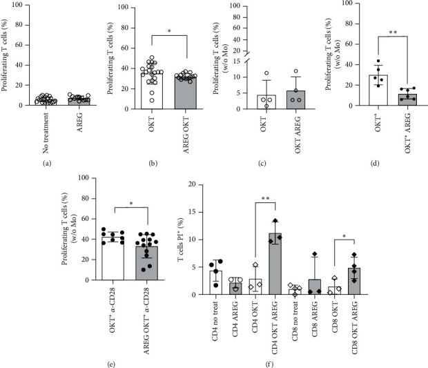
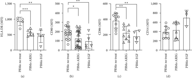
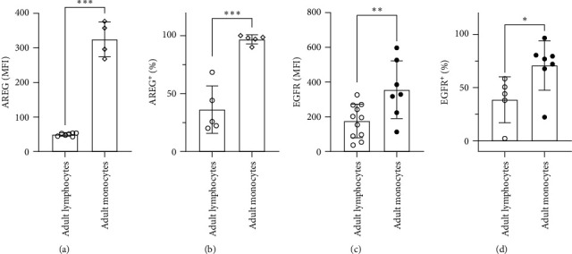
Amphiregulin (AREG) is a ligand of the epidermal growth factor receptor (EGFR) and has been shown to regulate the phagocytosis-induced cell death of monocytes in peripheral blood. AREG-dependent apoptotic signaling engages factors of the intrinsic and extrinsic apoptotic pathway, such as BCL-2, BCL-XL, and death ligand/receptor CD95/CD95L. Here, we tested the hypothesis that AREG influences costimulatory monocyte functions, which are crucial for T-cell responses. We found a stronger expression of AREG and EGFR in monocytes compared to lymphocytes. As a novel function of AREG, we observed reduced T-cell proliferation following polyclonal T-cell stimulation with OKT3. This reduction of proliferation occurred in the presence of monocytes as well as in their absence, monocyte signaling being replaced by crosslinking of OKT3. Increasing concentrations of AREG down-modulated the concentration of costimulatory B7 molecules (CD80/CD86) and HLA-DR on monocytes. In proliferation assays, CD28 expression on T cells was down-modulated on the application of OKT3 but unaltered by AREG. LcK activation, following OKT3-stimulation, was reduced in T cells that had been coincubated with AREG. The effects of AREG on T-cell phenotypes were also present when monocytes were depleted and OKT3 was crosslinked. The rearranged expression of immunological synapse proteins was accompanied by an alteration of T-cell polarization. Although the proportion of regulatory T cells was not shifted by AREG, IL-17-expressing T cells were significantly enhanced, with a bias toward TH1-polarization. Taken together, these results suggest that AREG acts as an immunoregulatory molecule at the interface between antigen-presenting cells and T cells.
期刊介绍:
Journal of Immunology Research is a peer-reviewed, Open Access journal that provides a platform for scientists and clinicians working in different areas of immunology and therapy. The journal publishes research articles, review articles, as well as clinical studies related to classical immunology, molecular immunology, clinical immunology, cancer immunology, transplantation immunology, immune pathology, immunodeficiency, autoimmune diseases, immune disorders, and immunotherapy.


 分享
分享
 求助内容:
求助内容: 应助结果提醒方式:
 扫码关注我们
