{"title":"18F-FDG PET/CT 在发现腹膜后副神经节瘤肺转移方面的效果。","authors":"Tomonori Chikasue, Seiji Kurata, Shuji Nagata, Shuichi Tanoue, Akiko Sumi, Mizuki Gobaru, Toru Hisaka, Toshihiro Hashiguchi, Takuya Furuta, Jun Akiba, Kiminori Fujimoto, Toshi Abe","doi":"10.22038/AOJNMB.2023.74066.1516","DOIUrl":null,"url":null,"abstract":"<p><p>A 50-year-old woman was diagnosed with iron deficiency anemia on general medical examination. Further, contrast-enhanced abdominal CT and magnetic resonance imaging revealed a large hypervascular mass with internal degeneration and necrosis in the retroperitoneal space. She was referred to our hospital for further evaluation and treatment. Because the paraganglioma was most likely as the imaging diagnosis, <sup>123</sup>I-MIBG scintigraphy was performed. It revealed the marked abnormal accumulation in the retroperitoneal lesion indicating the paraganglioma and no other abnormal accumulation was noted. Several plasma catecholamines and their urinary metabolites were normal. On the subsequent <sup>18</sup>F-FDG PET/CT, high FDG uptake was found in the retroperitoneal lesion (SUV<sub>max</sub>=38). FDG uptake was also found in a small nodule at the base of the lower lobe of the right lung (SUV<sub>max</sub>= 9.8). Contrast-enhanced imaging revealed a hypervascular nodule at the base of the right lung, suggesting pulmonary metastasis of a paraganglioma. The abdominal lesion and right lung nodule were excised, and retroperitoneal paraganglioma and pulmonary metastasis were diagnosed based on the pathology findings. In this case, <sup>18</sup>F-FDG PET/CT was useful in the search for paraganglioma metastasis. We report a relationship between <sup>123</sup>I-MIBG accumulation and <sup>18</sup>F-FDG uptake in paraganglioma and review the relevant literature.</p>","PeriodicalId":8503,"journal":{"name":"Asia Oceania Journal of Nuclear Medicine and Biology","volume":"12 1","pages":"46-51"},"PeriodicalIF":0.0000,"publicationDate":"2024-01-01","publicationTypes":"Journal Article","fieldsOfStudy":null,"isOpenAccess":false,"openAccessPdf":"https://www.ncbi.nlm.nih.gov/pmc/articles/PMC10757060/pdf/","citationCount":"0","resultStr":"{\"title\":\"Effectiveness of <sup>18</sup>F-FDG PET/CT in finding lung metastasis from a retroperitoneal paraganglioma.\",\"authors\":\"Tomonori Chikasue, Seiji Kurata, Shuji Nagata, Shuichi Tanoue, Akiko Sumi, Mizuki Gobaru, Toru Hisaka, Toshihiro Hashiguchi, Takuya Furuta, Jun Akiba, Kiminori Fujimoto, Toshi Abe\",\"doi\":\"10.22038/AOJNMB.2023.74066.1516\",\"DOIUrl\":null,\"url\":null,\"abstract\":\"<p><p>A 50-year-old woman was diagnosed with iron deficiency anemia on general medical examination. Further, contrast-enhanced abdominal CT and magnetic resonance imaging revealed a large hypervascular mass with internal degeneration and necrosis in the retroperitoneal space. She was referred to our hospital for further evaluation and treatment. Because the paraganglioma was most likely as the imaging diagnosis, <sup>123</sup>I-MIBG scintigraphy was performed. It revealed the marked abnormal accumulation in the retroperitoneal lesion indicating the paraganglioma and no other abnormal accumulation was noted. Several plasma catecholamines and their urinary metabolites were normal. On the subsequent <sup>18</sup>F-FDG PET/CT, high FDG uptake was found in the retroperitoneal lesion (SUV<sub>max</sub>=38). FDG uptake was also found in a small nodule at the base of the lower lobe of the right lung (SUV<sub>max</sub>= 9.8). Contrast-enhanced imaging revealed a hypervascular nodule at the base of the right lung, suggesting pulmonary metastasis of a paraganglioma. The abdominal lesion and right lung nodule were excised, and retroperitoneal paraganglioma and pulmonary metastasis were diagnosed based on the pathology findings. In this case, <sup>18</sup>F-FDG PET/CT was useful in the search for paraganglioma metastasis. We report a relationship between <sup>123</sup>I-MIBG accumulation and <sup>18</sup>F-FDG uptake in paraganglioma and review the relevant literature.</p>\",\"PeriodicalId\":8503,\"journal\":{\"name\":\"Asia Oceania Journal of Nuclear Medicine and Biology\",\"volume\":\"12 1\",\"pages\":\"46-51\"},\"PeriodicalIF\":0.0000,\"publicationDate\":\"2024-01-01\",\"publicationTypes\":\"Journal Article\",\"fieldsOfStudy\":null,\"isOpenAccess\":false,\"openAccessPdf\":\"https://www.ncbi.nlm.nih.gov/pmc/articles/PMC10757060/pdf/\",\"citationCount\":\"0\",\"resultStr\":null,\"platform\":\"Semanticscholar\",\"paperid\":null,\"PeriodicalName\":\"Asia Oceania Journal of Nuclear Medicine and Biology\",\"FirstCategoryId\":\"1085\",\"ListUrlMain\":\"https://doi.org/10.22038/AOJNMB.2023.74066.1516\",\"RegionNum\":0,\"RegionCategory\":null,\"ArticlePicture\":[],\"TitleCN\":null,\"AbstractTextCN\":null,\"PMCID\":null,\"EPubDate\":\"\",\"PubModel\":\"\",\"JCR\":\"Q3\",\"JCRName\":\"Medicine\",\"Score\":null,\"Total\":0}","platform":"Semanticscholar","paperid":null,"PeriodicalName":"Asia Oceania Journal of Nuclear Medicine and Biology","FirstCategoryId":"1085","ListUrlMain":"https://doi.org/10.22038/AOJNMB.2023.74066.1516","RegionNum":0,"RegionCategory":null,"ArticlePicture":[],"TitleCN":null,"AbstractTextCN":null,"PMCID":null,"EPubDate":"","PubModel":"","JCR":"Q3","JCRName":"Medicine","Score":null,"Total":0}
Effectiveness of 18F-FDG PET/CT in finding lung metastasis from a retroperitoneal paraganglioma.
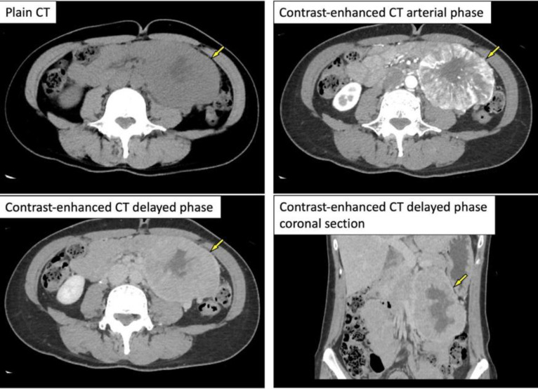
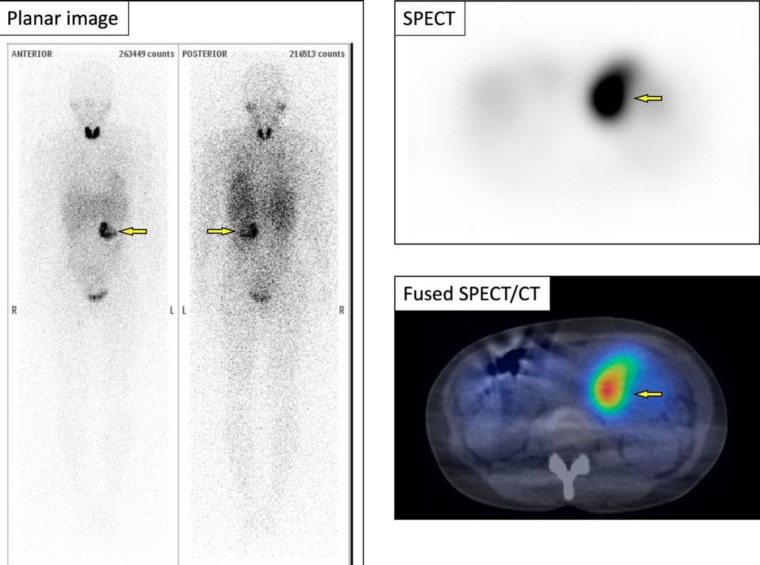
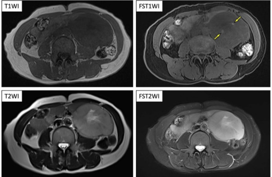
A 50-year-old woman was diagnosed with iron deficiency anemia on general medical examination. Further, contrast-enhanced abdominal CT and magnetic resonance imaging revealed a large hypervascular mass with internal degeneration and necrosis in the retroperitoneal space. She was referred to our hospital for further evaluation and treatment. Because the paraganglioma was most likely as the imaging diagnosis, 123I-MIBG scintigraphy was performed. It revealed the marked abnormal accumulation in the retroperitoneal lesion indicating the paraganglioma and no other abnormal accumulation was noted. Several plasma catecholamines and their urinary metabolites were normal. On the subsequent 18F-FDG PET/CT, high FDG uptake was found in the retroperitoneal lesion (SUVmax=38). FDG uptake was also found in a small nodule at the base of the lower lobe of the right lung (SUVmax= 9.8). Contrast-enhanced imaging revealed a hypervascular nodule at the base of the right lung, suggesting pulmonary metastasis of a paraganglioma. The abdominal lesion and right lung nodule were excised, and retroperitoneal paraganglioma and pulmonary metastasis were diagnosed based on the pathology findings. In this case, 18F-FDG PET/CT was useful in the search for paraganglioma metastasis. We report a relationship between 123I-MIBG accumulation and 18F-FDG uptake in paraganglioma and review the relevant literature.


 分享
分享
 求助内容:
求助内容: 应助结果提醒方式:
 扫码关注我们
