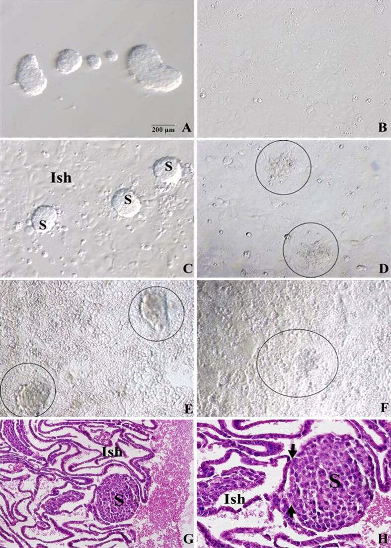
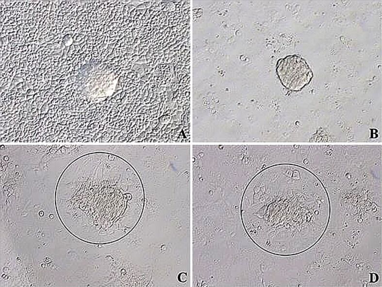
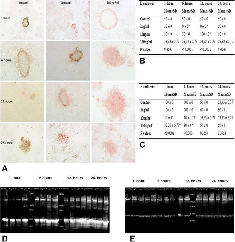
{"title":"肝素结合表皮生长因子对植入粘附阶段的体外影响。","authors":"Burcu Biltekin, Ayhan Bilir, Ismail Seckin, Gozde Erkanli Senturk","doi":"10.47162/RJME.64.4.05","DOIUrl":null,"url":null,"abstract":"<p><p>A member of the epidermal growth factor (EGF) family, the heparin-binding EGF (HB-EGF) is expressed in the uteri of both humans and mice during the implantation process. To study the effects of HB-EGF on adhesion stage, we developed an in vitro implantation model employing Ishikawa cell line and JAR cell line, which may attach to Ishikawa cells. For 1, 6, 12, and 24 hours, co-cultures of JAR spheroids grown on Ishikawa monolayers were treated with 1, 10, and 100 ng∕mL doses of HB-EGF. Using immunocytochemistry and Western blot analysis, the effects of HB-EGF on the protein expressions of E-cadherin, Erb-B2 receptor tyrosine kinase 4 (ErbB4), and integrin ανβ3 in Ishikawa and JAR cells were examined semi-quantitatively and quantitatively. Ultrastructural changes of in vitro implantation model were investigated by transmission electron microscopy. We revealed that HB-EGF influenced trophoblast cell adhesion to endometrial cells by upregulating the expression of the proteins ErbB4 and trophoblastic integrin ανβ3. Decrease in trophoblastic E-cadherin expression and increase in endometrial E-cadherin expression were demonstrated accompanying morphological variations in cells required for the invasion. We discovered ultrastructurally that Ishikawa cells acquired uterodome-like appearance, including the organelles, when 10 and 100 ng∕mL dosages of HB-EGF were administered for 12 and 24 hours. However, following additional hours of adhesion and invasion, their intercellular spaces enlarged. The trafficking of vesicular transport was enhanced by JAR spheroids. We therefore discovered that in this implantation paradigm, HB-EGF may enhance the receptivity of Ishikawa cells and the adherence of JAR cells.</p>","PeriodicalId":54447,"journal":{"name":"Romanian Journal of Morphology and Embryology","volume":"64 4","pages":"493-500"},"PeriodicalIF":1.5000,"publicationDate":"2023-10-01","publicationTypes":"Journal Article","fieldsOfStudy":null,"isOpenAccess":false,"openAccessPdf":"https://www.ncbi.nlm.nih.gov/pmc/articles/PMC10863694/pdf/","citationCount":"0","resultStr":"{\"title\":\"In vitro effects of heparin-binding epidermal growth factor on adhesion stage of implantation.\",\"authors\":\"Burcu Biltekin, Ayhan Bilir, Ismail Seckin, Gozde Erkanli Senturk\",\"doi\":\"10.47162/RJME.64.4.05\",\"DOIUrl\":null,\"url\":null,\"abstract\":\"<p><p>A member of the epidermal growth factor (EGF) family, the heparin-binding EGF (HB-EGF) is expressed in the uteri of both humans and mice during the implantation process. To study the effects of HB-EGF on adhesion stage, we developed an in vitro implantation model employing Ishikawa cell line and JAR cell line, which may attach to Ishikawa cells. For 1, 6, 12, and 24 hours, co-cultures of JAR spheroids grown on Ishikawa monolayers were treated with 1, 10, and 100 ng∕mL doses of HB-EGF. Using immunocytochemistry and Western blot analysis, the effects of HB-EGF on the protein expressions of E-cadherin, Erb-B2 receptor tyrosine kinase 4 (ErbB4), and integrin ανβ3 in Ishikawa and JAR cells were examined semi-quantitatively and quantitatively. Ultrastructural changes of in vitro implantation model were investigated by transmission electron microscopy. We revealed that HB-EGF influenced trophoblast cell adhesion to endometrial cells by upregulating the expression of the proteins ErbB4 and trophoblastic integrin ανβ3. Decrease in trophoblastic E-cadherin expression and increase in endometrial E-cadherin expression were demonstrated accompanying morphological variations in cells required for the invasion. We discovered ultrastructurally that Ishikawa cells acquired uterodome-like appearance, including the organelles, when 10 and 100 ng∕mL dosages of HB-EGF were administered for 12 and 24 hours. However, following additional hours of adhesion and invasion, their intercellular spaces enlarged. The trafficking of vesicular transport was enhanced by JAR spheroids. We therefore discovered that in this implantation paradigm, HB-EGF may enhance the receptivity of Ishikawa cells and the adherence of JAR cells.</p>\",\"PeriodicalId\":54447,\"journal\":{\"name\":\"Romanian Journal of Morphology and Embryology\",\"volume\":\"64 4\",\"pages\":\"493-500\"},\"PeriodicalIF\":1.5000,\"publicationDate\":\"2023-10-01\",\"publicationTypes\":\"Journal Article\",\"fieldsOfStudy\":null,\"isOpenAccess\":false,\"openAccessPdf\":\"https://www.ncbi.nlm.nih.gov/pmc/articles/PMC10863694/pdf/\",\"citationCount\":\"0\",\"resultStr\":null,\"platform\":\"Semanticscholar\",\"paperid\":null,\"PeriodicalName\":\"Romanian Journal of Morphology and Embryology\",\"FirstCategoryId\":\"3\",\"ListUrlMain\":\"https://doi.org/10.47162/RJME.64.4.05\",\"RegionNum\":4,\"RegionCategory\":\"医学\",\"ArticlePicture\":[],\"TitleCN\":null,\"AbstractTextCN\":null,\"PMCID\":null,\"EPubDate\":\"\",\"PubModel\":\"\",\"JCR\":\"Q4\",\"JCRName\":\"DEVELOPMENTAL BIOLOGY\",\"Score\":null,\"Total\":0}","platform":"Semanticscholar","paperid":null,"PeriodicalName":"Romanian Journal of Morphology and Embryology","FirstCategoryId":"3","ListUrlMain":"https://doi.org/10.47162/RJME.64.4.05","RegionNum":4,"RegionCategory":"医学","ArticlePicture":[],"TitleCN":null,"AbstractTextCN":null,"PMCID":null,"EPubDate":"","PubModel":"","JCR":"Q4","JCRName":"DEVELOPMENTAL BIOLOGY","Score":null,"Total":0}
引用次数: 0
摘要
作为表皮生长因子(EGF)家族的一员,肝素结合型 EGF(HB-EGF)在人类和小鼠的子宫植入过程中均有表达。为了研究 HB-EGF 对粘附阶段的影响,我们利用石川细胞系和 JAR 细胞系建立了一个体外植入模型。在1、6、12和24小时内,用1、10和100 ng∕mL剂量的HB-EGF处理在石川单层细胞上生长的JAR球状细胞的共培养物。通过免疫细胞化学和 Western 印迹分析,半定量和定量分析了 HB-EGF 对石川细胞和 JAR 细胞中 E-钙粘连蛋白、Erb-B2 受体酪氨酸激酶 4(ErbB4)和整合素 ανβ3 蛋白表达的影响。透射电子显微镜研究了体外植入模型的超微结构变化。我们发现,HB-EGF通过上调ErbB4蛋白和滋养层整合素ανβ3的表达影响滋养层细胞与子宫内膜细胞的粘附。滋养细胞E-cadherin表达的减少和子宫内膜E-cadherin表达的增加伴随着入侵所需的细胞形态变化。我们在超微结构上发现,当给予 10 和 100 ng∕mL 剂量的 HB-EGF 12 和 24 小时后,石川细胞获得了子宫瘤样外观,包括细胞器。然而,经过数小时的粘附和侵袭后,它们的细胞间隙扩大了。JAR球体增强了囊泡运输。因此,我们发现在这种植入模式中,HB-EGF 可增强石川细胞的接受能力和 JAR 细胞的粘附能力。
In vitro effects of heparin-binding epidermal growth factor on adhesion stage of implantation.
A member of the epidermal growth factor (EGF) family, the heparin-binding EGF (HB-EGF) is expressed in the uteri of both humans and mice during the implantation process. To study the effects of HB-EGF on adhesion stage, we developed an in vitro implantation model employing Ishikawa cell line and JAR cell line, which may attach to Ishikawa cells. For 1, 6, 12, and 24 hours, co-cultures of JAR spheroids grown on Ishikawa monolayers were treated with 1, 10, and 100 ng∕mL doses of HB-EGF. Using immunocytochemistry and Western blot analysis, the effects of HB-EGF on the protein expressions of E-cadherin, Erb-B2 receptor tyrosine kinase 4 (ErbB4), and integrin ανβ3 in Ishikawa and JAR cells were examined semi-quantitatively and quantitatively. Ultrastructural changes of in vitro implantation model were investigated by transmission electron microscopy. We revealed that HB-EGF influenced trophoblast cell adhesion to endometrial cells by upregulating the expression of the proteins ErbB4 and trophoblastic integrin ανβ3. Decrease in trophoblastic E-cadherin expression and increase in endometrial E-cadherin expression were demonstrated accompanying morphological variations in cells required for the invasion. We discovered ultrastructurally that Ishikawa cells acquired uterodome-like appearance, including the organelles, when 10 and 100 ng∕mL dosages of HB-EGF were administered for 12 and 24 hours. However, following additional hours of adhesion and invasion, their intercellular spaces enlarged. The trafficking of vesicular transport was enhanced by JAR spheroids. We therefore discovered that in this implantation paradigm, HB-EGF may enhance the receptivity of Ishikawa cells and the adherence of JAR cells.
期刊介绍:
Romanian Journal of Morphology and Embryology (Rom J Morphol Embryol) publishes studies on all aspects of normal morphology and human comparative and experimental pathology. The Journal accepts only researches that utilize modern investigation methods (studies of anatomy, pathology, cytopathology, immunohistochemistry, histochemistry, immunology, morphometry, molecular and cellular biology, electronic microscopy, etc.).


 分享
分享
 求助内容:
求助内容: 应助结果提醒方式:
 扫码关注我们
