Banu Farabi, Mehmet Fatih Atak, Ucalene Harris, Julia Kahn, Samavia Khan, Veronica Fink, Daniella Hartmann, Babar K. Rao, Manu Jain
{"title":"模拟非黑色素瘤皮肤癌的常见良性病变的体外共聚焦显微镜特征:实现临床整合。","authors":"Banu Farabi, Mehmet Fatih Atak, Ucalene Harris, Julia Kahn, Samavia Khan, Veronica Fink, Daniella Hartmann, Babar K. Rao, Manu Jain","doi":"10.1002/jbio.202300386","DOIUrl":null,"url":null,"abstract":"<p>Ex vivo confocal microscope (EVCM) rapidly images freshly excised tissue at a histopathological resolution. EVCM features of keratinocyte skin cancers are well-established, but those of benign clinical mimickers remain scarce. We describe EVCM features of common benign lesions and compare them with their malignant differentials. EVCM was used to image 14 benign and 3 cancer tissues. We compared EVCM features of benign lesions with corresponding histopathology and with those of keratinocyte cancers. Key features of benign lesions were identified and differentiated from malignant lesions. Elastin and fat appeared prominent in EVCM; while koilocytes and melanin were difficult to identify. Visualization of entire epidermis was challenging due to difficulty of tissue flattening during imaging. Benign lesions can be differentiated from keratinocyte cancers with EVCM. Using EVCM, a rapid, bedside diagnosis and management of skin neoplasms is possible, especially in a remote location without a histopathology lab.</p>","PeriodicalId":184,"journal":{"name":"Journal of Biophotonics","volume":"17 4","pages":""},"PeriodicalIF":2.0000,"publicationDate":"2024-01-10","publicationTypes":"Journal Article","fieldsOfStudy":null,"isOpenAccess":false,"openAccessPdf":"","citationCount":"0","resultStr":"{\"title\":\"Ex vivo confocal microscopy features of common benign lesions that mimic non-melanoma skin cancers: Towards clinical integration\",\"authors\":\"Banu Farabi, Mehmet Fatih Atak, Ucalene Harris, Julia Kahn, Samavia Khan, Veronica Fink, Daniella Hartmann, Babar K. Rao, Manu Jain\",\"doi\":\"10.1002/jbio.202300386\",\"DOIUrl\":null,\"url\":null,\"abstract\":\"<p>Ex vivo confocal microscope (EVCM) rapidly images freshly excised tissue at a histopathological resolution. EVCM features of keratinocyte skin cancers are well-established, but those of benign clinical mimickers remain scarce. We describe EVCM features of common benign lesions and compare them with their malignant differentials. EVCM was used to image 14 benign and 3 cancer tissues. We compared EVCM features of benign lesions with corresponding histopathology and with those of keratinocyte cancers. Key features of benign lesions were identified and differentiated from malignant lesions. Elastin and fat appeared prominent in EVCM; while koilocytes and melanin were difficult to identify. Visualization of entire epidermis was challenging due to difficulty of tissue flattening during imaging. Benign lesions can be differentiated from keratinocyte cancers with EVCM. Using EVCM, a rapid, bedside diagnosis and management of skin neoplasms is possible, especially in a remote location without a histopathology lab.</p>\",\"PeriodicalId\":184,\"journal\":{\"name\":\"Journal of Biophotonics\",\"volume\":\"17 4\",\"pages\":\"\"},\"PeriodicalIF\":2.0000,\"publicationDate\":\"2024-01-10\",\"publicationTypes\":\"Journal Article\",\"fieldsOfStudy\":null,\"isOpenAccess\":false,\"openAccessPdf\":\"\",\"citationCount\":\"0\",\"resultStr\":null,\"platform\":\"Semanticscholar\",\"paperid\":null,\"PeriodicalName\":\"Journal of Biophotonics\",\"FirstCategoryId\":\"101\",\"ListUrlMain\":\"https://onlinelibrary.wiley.com/doi/10.1002/jbio.202300386\",\"RegionNum\":3,\"RegionCategory\":\"物理与天体物理\",\"ArticlePicture\":[],\"TitleCN\":null,\"AbstractTextCN\":null,\"PMCID\":null,\"EPubDate\":\"\",\"PubModel\":\"\",\"JCR\":\"Q3\",\"JCRName\":\"BIOCHEMICAL RESEARCH METHODS\",\"Score\":null,\"Total\":0}","platform":"Semanticscholar","paperid":null,"PeriodicalName":"Journal of Biophotonics","FirstCategoryId":"101","ListUrlMain":"https://onlinelibrary.wiley.com/doi/10.1002/jbio.202300386","RegionNum":3,"RegionCategory":"物理与天体物理","ArticlePicture":[],"TitleCN":null,"AbstractTextCN":null,"PMCID":null,"EPubDate":"","PubModel":"","JCR":"Q3","JCRName":"BIOCHEMICAL RESEARCH METHODS","Score":null,"Total":0}
Ex vivo confocal microscopy features of common benign lesions that mimic non-melanoma skin cancers: Towards clinical integration
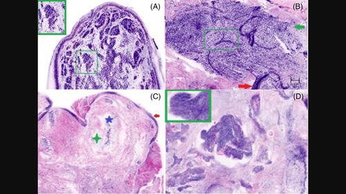
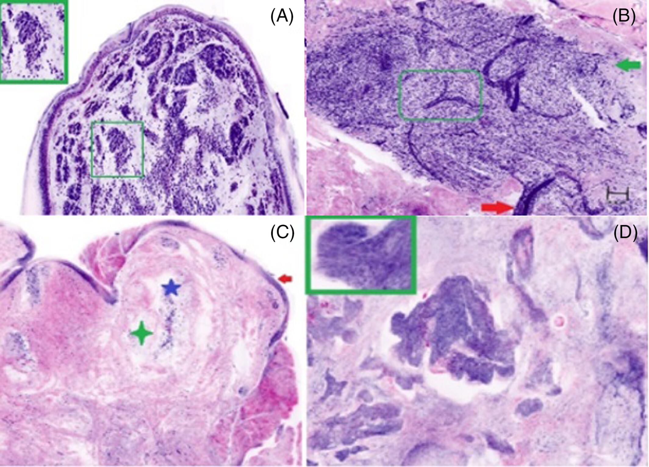
Ex vivo confocal microscope (EVCM) rapidly images freshly excised tissue at a histopathological resolution. EVCM features of keratinocyte skin cancers are well-established, but those of benign clinical mimickers remain scarce. We describe EVCM features of common benign lesions and compare them with their malignant differentials. EVCM was used to image 14 benign and 3 cancer tissues. We compared EVCM features of benign lesions with corresponding histopathology and with those of keratinocyte cancers. Key features of benign lesions were identified and differentiated from malignant lesions. Elastin and fat appeared prominent in EVCM; while koilocytes and melanin were difficult to identify. Visualization of entire epidermis was challenging due to difficulty of tissue flattening during imaging. Benign lesions can be differentiated from keratinocyte cancers with EVCM. Using EVCM, a rapid, bedside diagnosis and management of skin neoplasms is possible, especially in a remote location without a histopathology lab.
期刊介绍:
The first international journal dedicated to publishing reviews and original articles from this exciting field, the Journal of Biophotonics covers the broad range of research on interactions between light and biological material. The journal offers a platform where the physicist communicates with the biologist and where the clinical practitioner learns about the latest tools for the diagnosis of diseases. As such, the journal is highly interdisciplinary, publishing cutting edge research in the fields of life sciences, medicine, physics, chemistry, and engineering. The coverage extends from fundamental research to specific developments, while also including the latest applications.


 分享
分享
 求助内容:
求助内容: 应助结果提醒方式:
 扫码关注我们
