M. Manuela Brás, Aureliana Sousa, Tânia B. Cruz, Jonas Michalewski, Marina Leite, Susana R. Sousa, Pedro L. Granja, Manfred Radmacher
{"title":"利用原子力显微镜对黑色素瘤细胞进行微流变学比较。","authors":"M. Manuela Brás, Aureliana Sousa, Tânia B. Cruz, Jonas Michalewski, Marina Leite, Susana R. Sousa, Pedro L. Granja, Manfred Radmacher","doi":"10.1007/s10867-023-09648-w","DOIUrl":null,"url":null,"abstract":"<div><p>Melanoma is one of the most severe cancers due to its great potential to form metastasis. Recent studies showed the importance of mechanical property assessment in metastasis formation which depends on the cytoskeleton dynamics and cell migration. Although cells are considered purely elastic, they are viscoelastic entities. Microrheology atomic force microscopy (AFM) enables the assessment of elasticity and viscous properties, which are relevant to cell behavior regulation. The current work compares the mechanical properties of human neonatal primary melanocytes (HNPMs) with two melanoma cell lines (WM793B and 1205LU cells), using microrheology AFM. Immunocytochemistry of F-actin filaments and phosphorylated focal adhesion kinase (p-FAK) and cell migration assays were performed to understand the differences found in microrheology AFM regarding the tumor cell lines tested. AFM revealed that HNPMs and tumor cell lines had distinct mechanical properties. HNPMs were softer, less viscous, presenting a higher power-law than melanoma cells. Immunostaining showed that metastatic 1205LU cells expressed more p-FAK than WM793B cells. Melanoma cell migration assays showed that WM73B did not close the gap, in contrast to 1205LU cells, which closed the gap at the end of 23 h. These data seem to corroborate the high migratory behavior of 1205LU cells. Microrheology AFM applied to HNPMs and melanoma cells allowed the quantification of elasticity, viscous properties, glassy phase, and power-law properties, which have an impact in cell migration and metastasis formation. AFM study is important since it can be used as a biomarker of the different stages of the disease in melanoma.</p></div>","PeriodicalId":612,"journal":{"name":"Journal of Biological Physics","volume":"50 1","pages":"55 - 69"},"PeriodicalIF":2.0000,"publicationDate":"2024-01-19","publicationTypes":"Journal Article","fieldsOfStudy":null,"isOpenAccess":false,"openAccessPdf":"https://www.ncbi.nlm.nih.gov/pmc/articles/PMC10864228/pdf/","citationCount":"0","resultStr":"{\"title\":\"Microrheological comparison of melanoma cells by atomic force microscopy\",\"authors\":\"M. Manuela Brás, Aureliana Sousa, Tânia B. Cruz, Jonas Michalewski, Marina Leite, Susana R. Sousa, Pedro L. Granja, Manfred Radmacher\",\"doi\":\"10.1007/s10867-023-09648-w\",\"DOIUrl\":null,\"url\":null,\"abstract\":\"<div><p>Melanoma is one of the most severe cancers due to its great potential to form metastasis. Recent studies showed the importance of mechanical property assessment in metastasis formation which depends on the cytoskeleton dynamics and cell migration. Although cells are considered purely elastic, they are viscoelastic entities. Microrheology atomic force microscopy (AFM) enables the assessment of elasticity and viscous properties, which are relevant to cell behavior regulation. The current work compares the mechanical properties of human neonatal primary melanocytes (HNPMs) with two melanoma cell lines (WM793B and 1205LU cells), using microrheology AFM. Immunocytochemistry of F-actin filaments and phosphorylated focal adhesion kinase (p-FAK) and cell migration assays were performed to understand the differences found in microrheology AFM regarding the tumor cell lines tested. AFM revealed that HNPMs and tumor cell lines had distinct mechanical properties. HNPMs were softer, less viscous, presenting a higher power-law than melanoma cells. Immunostaining showed that metastatic 1205LU cells expressed more p-FAK than WM793B cells. Melanoma cell migration assays showed that WM73B did not close the gap, in contrast to 1205LU cells, which closed the gap at the end of 23 h. These data seem to corroborate the high migratory behavior of 1205LU cells. Microrheology AFM applied to HNPMs and melanoma cells allowed the quantification of elasticity, viscous properties, glassy phase, and power-law properties, which have an impact in cell migration and metastasis formation. AFM study is important since it can be used as a biomarker of the different stages of the disease in melanoma.</p></div>\",\"PeriodicalId\":612,\"journal\":{\"name\":\"Journal of Biological Physics\",\"volume\":\"50 1\",\"pages\":\"55 - 69\"},\"PeriodicalIF\":2.0000,\"publicationDate\":\"2024-01-19\",\"publicationTypes\":\"Journal Article\",\"fieldsOfStudy\":null,\"isOpenAccess\":false,\"openAccessPdf\":\"https://www.ncbi.nlm.nih.gov/pmc/articles/PMC10864228/pdf/\",\"citationCount\":\"0\",\"resultStr\":null,\"platform\":\"Semanticscholar\",\"paperid\":null,\"PeriodicalName\":\"Journal of Biological Physics\",\"FirstCategoryId\":\"99\",\"ListUrlMain\":\"https://link.springer.com/article/10.1007/s10867-023-09648-w\",\"RegionNum\":4,\"RegionCategory\":\"生物学\",\"ArticlePicture\":[],\"TitleCN\":null,\"AbstractTextCN\":null,\"PMCID\":null,\"EPubDate\":\"\",\"PubModel\":\"\",\"JCR\":\"Q3\",\"JCRName\":\"BIOPHYSICS\",\"Score\":null,\"Total\":0}","platform":"Semanticscholar","paperid":null,"PeriodicalName":"Journal of Biological Physics","FirstCategoryId":"99","ListUrlMain":"https://link.springer.com/article/10.1007/s10867-023-09648-w","RegionNum":4,"RegionCategory":"生物学","ArticlePicture":[],"TitleCN":null,"AbstractTextCN":null,"PMCID":null,"EPubDate":"","PubModel":"","JCR":"Q3","JCRName":"BIOPHYSICS","Score":null,"Total":0}
Microrheological comparison of melanoma cells by atomic force microscopy
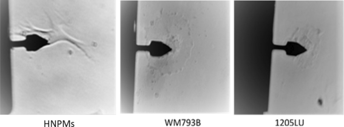
Melanoma is one of the most severe cancers due to its great potential to form metastasis. Recent studies showed the importance of mechanical property assessment in metastasis formation which depends on the cytoskeleton dynamics and cell migration. Although cells are considered purely elastic, they are viscoelastic entities. Microrheology atomic force microscopy (AFM) enables the assessment of elasticity and viscous properties, which are relevant to cell behavior regulation. The current work compares the mechanical properties of human neonatal primary melanocytes (HNPMs) with two melanoma cell lines (WM793B and 1205LU cells), using microrheology AFM. Immunocytochemistry of F-actin filaments and phosphorylated focal adhesion kinase (p-FAK) and cell migration assays were performed to understand the differences found in microrheology AFM regarding the tumor cell lines tested. AFM revealed that HNPMs and tumor cell lines had distinct mechanical properties. HNPMs were softer, less viscous, presenting a higher power-law than melanoma cells. Immunostaining showed that metastatic 1205LU cells expressed more p-FAK than WM793B cells. Melanoma cell migration assays showed that WM73B did not close the gap, in contrast to 1205LU cells, which closed the gap at the end of 23 h. These data seem to corroborate the high migratory behavior of 1205LU cells. Microrheology AFM applied to HNPMs and melanoma cells allowed the quantification of elasticity, viscous properties, glassy phase, and power-law properties, which have an impact in cell migration and metastasis formation. AFM study is important since it can be used as a biomarker of the different stages of the disease in melanoma.
期刊介绍:
Many physicists are turning their attention to domains that were not traditionally part of physics and are applying the sophisticated tools of theoretical, computational and experimental physics to investigate biological processes, systems and materials.
The Journal of Biological Physics provides a medium where this growing community of scientists can publish its results and discuss its aims and methods. It welcomes papers which use the tools of physics in an innovative way to study biological problems, as well as research aimed at providing a better understanding of the physical principles underlying biological processes.


 分享
分享
 求助内容:
求助内容: 应助结果提醒方式:
 扫码关注我们
