Daniele Antonio Pizzuto, Lucio Calandriello, Ivan De Martino, Maria Luisa De Micheli, Marco De Summa, Salvatore Annunziata
{"title":"肌肉骨骼疾病中的正电子发射断层扫描/磁共振:正确的序列和工作流程优化","authors":"Daniele Antonio Pizzuto, Lucio Calandriello, Ivan De Martino, Maria Luisa De Micheli, Marco De Summa, Salvatore Annunziata","doi":"10.1007/s40336-023-00611-2","DOIUrl":null,"url":null,"abstract":"<p>Magnetic resonance imaging (MRI) represents a gold standard imaging for detection of oncologic and non-oncologic musculoskeletal disorders (MSK), owing to its high soft tissue contrast. Positron Emission Tomography (PET) was proven to be clinically useful in MSK, owing to its early detection of metabolic disfunction and its high accuracy for monitoring therapy response. With hybrid PET/MRI system, simultaneous availability of both morphologic and metabolic features could potentially enhance the diagnostic accuracy in MSK. Some technical issue should be overcome for best imaging quality: specific MR sequences for accurate visualization of cortical bone and bone marrow involvement, such as zero-time echo (ZTE) or µ time echo (µTE) sequences, that were shown to provide valuable attenuation coefficients for the bone, which leads to accurate quantitative analysis of bone and extra-bone tissues; implementation of novel attenuation map, owing to the presence of flexible coils in the field of view, additional sequences to reduce artifacts derived from metal implants. Workflow consideration should be addressed to the choice of proper sequences able to answer the clinical demand or the research purpose. Redundant information provided by useless sequences, which could prolong the whole scan time and increase the discomfort of the patient, should be avoided.</p>","PeriodicalId":48600,"journal":{"name":"Clinical and Translational Imaging","volume":"59 1","pages":""},"PeriodicalIF":1.6000,"publicationDate":"2024-01-17","publicationTypes":"Journal Article","fieldsOfStudy":null,"isOpenAccess":false,"openAccessPdf":"","citationCount":"0","resultStr":"{\"title\":\"Positron emission tomography/magnetic resonance in musculoskeletal disorders: proper sequences and workflow optimization\",\"authors\":\"Daniele Antonio Pizzuto, Lucio Calandriello, Ivan De Martino, Maria Luisa De Micheli, Marco De Summa, Salvatore Annunziata\",\"doi\":\"10.1007/s40336-023-00611-2\",\"DOIUrl\":null,\"url\":null,\"abstract\":\"<p>Magnetic resonance imaging (MRI) represents a gold standard imaging for detection of oncologic and non-oncologic musculoskeletal disorders (MSK), owing to its high soft tissue contrast. Positron Emission Tomography (PET) was proven to be clinically useful in MSK, owing to its early detection of metabolic disfunction and its high accuracy for monitoring therapy response. With hybrid PET/MRI system, simultaneous availability of both morphologic and metabolic features could potentially enhance the diagnostic accuracy in MSK. Some technical issue should be overcome for best imaging quality: specific MR sequences for accurate visualization of cortical bone and bone marrow involvement, such as zero-time echo (ZTE) or µ time echo (µTE) sequences, that were shown to provide valuable attenuation coefficients for the bone, which leads to accurate quantitative analysis of bone and extra-bone tissues; implementation of novel attenuation map, owing to the presence of flexible coils in the field of view, additional sequences to reduce artifacts derived from metal implants. Workflow consideration should be addressed to the choice of proper sequences able to answer the clinical demand or the research purpose. Redundant information provided by useless sequences, which could prolong the whole scan time and increase the discomfort of the patient, should be avoided.</p>\",\"PeriodicalId\":48600,\"journal\":{\"name\":\"Clinical and Translational Imaging\",\"volume\":\"59 1\",\"pages\":\"\"},\"PeriodicalIF\":1.6000,\"publicationDate\":\"2024-01-17\",\"publicationTypes\":\"Journal Article\",\"fieldsOfStudy\":null,\"isOpenAccess\":false,\"openAccessPdf\":\"\",\"citationCount\":\"0\",\"resultStr\":null,\"platform\":\"Semanticscholar\",\"paperid\":null,\"PeriodicalName\":\"Clinical and Translational Imaging\",\"FirstCategoryId\":\"3\",\"ListUrlMain\":\"https://doi.org/10.1007/s40336-023-00611-2\",\"RegionNum\":4,\"RegionCategory\":\"医学\",\"ArticlePicture\":[],\"TitleCN\":null,\"AbstractTextCN\":null,\"PMCID\":null,\"EPubDate\":\"\",\"PubModel\":\"\",\"JCR\":\"Q2\",\"JCRName\":\"RADIOLOGY, NUCLEAR MEDICINE & MEDICAL IMAGING\",\"Score\":null,\"Total\":0}","platform":"Semanticscholar","paperid":null,"PeriodicalName":"Clinical and Translational Imaging","FirstCategoryId":"3","ListUrlMain":"https://doi.org/10.1007/s40336-023-00611-2","RegionNum":4,"RegionCategory":"医学","ArticlePicture":[],"TitleCN":null,"AbstractTextCN":null,"PMCID":null,"EPubDate":"","PubModel":"","JCR":"Q2","JCRName":"RADIOLOGY, NUCLEAR MEDICINE & MEDICAL IMAGING","Score":null,"Total":0}
Positron emission tomography/magnetic resonance in musculoskeletal disorders: proper sequences and workflow optimization
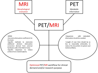
Magnetic resonance imaging (MRI) represents a gold standard imaging for detection of oncologic and non-oncologic musculoskeletal disorders (MSK), owing to its high soft tissue contrast. Positron Emission Tomography (PET) was proven to be clinically useful in MSK, owing to its early detection of metabolic disfunction and its high accuracy for monitoring therapy response. With hybrid PET/MRI system, simultaneous availability of both morphologic and metabolic features could potentially enhance the diagnostic accuracy in MSK. Some technical issue should be overcome for best imaging quality: specific MR sequences for accurate visualization of cortical bone and bone marrow involvement, such as zero-time echo (ZTE) or µ time echo (µTE) sequences, that were shown to provide valuable attenuation coefficients for the bone, which leads to accurate quantitative analysis of bone and extra-bone tissues; implementation of novel attenuation map, owing to the presence of flexible coils in the field of view, additional sequences to reduce artifacts derived from metal implants. Workflow consideration should be addressed to the choice of proper sequences able to answer the clinical demand or the research purpose. Redundant information provided by useless sequences, which could prolong the whole scan time and increase the discomfort of the patient, should be avoided.
期刊介绍:
Clinical and Translational Imaging is an international journal that publishes timely, up-to-date summaries on clinical practice and translational research and clinical applications of approved and experimental radiopharmaceuticals for diagnostic and therapeutic purposes. Coverage includes such topics as advanced preclinical evidence in the fields of physics, dosimetry, radiation biology and radiopharmacy with relevance to applications in human subjects. The journal benefits a readership of nuclear medicine practitioners and allied professionals involved in molecular imaging and therapy.


 分享
分享
 求助内容:
求助内容: 应助结果提醒方式:
 扫码关注我们
