Megan Clapperton, Tash Kunanandam, Catalina D. Florea, Catriona M. Douglas, Gail McConnell
{"title":"多模态光学介孔镜揭示了体外扁桃体中革兰氏阳性生物膜的数量和空间分布。","authors":"Megan Clapperton, Tash Kunanandam, Catalina D. Florea, Catriona M. Douglas, Gail McConnell","doi":"10.1111/jmi.13266","DOIUrl":null,"url":null,"abstract":"<p>Biofilms are known to be present in tonsils, but little is known about their spatial location and size distribution throughout the tonsil. Studies of the location and distribution of biofilms in tonsil specimens have thus far been limited to either high-magnification methods such as electron microscopy, which enables high-resolution imaging but only from a tiny tissue volume, or lower magnification techniques such as light microscopy, which allow imaging of larger specimens but with poor spatial resolution. To overcome these limitations, we report the use of multimodal optical mesoscopy to visualise and quantify the number and spatial distribution of Gram-positive biofilms in fresh, excised paediatric tonsils. This methodology supports simultaneous imaging of both the tonsil host and biofilms in whole mounts of tissue up to 5 mm × 5 mm × 3 mm with subcellular resolution throughout. A quantitative assessment of 36 tonsil specimens revealed no statistically significant difference between biofilm presence on the tonsil surface and the interior of the tonsil. This new quantitative mesoscale imaging approach may prove useful in understanding the role of biofilms in tonsillar diseases and other infections.</p>","PeriodicalId":16484,"journal":{"name":"Journal of microscopy","volume":"295 2","pages":"121-130"},"PeriodicalIF":1.9000,"publicationDate":"2024-01-31","publicationTypes":"Journal Article","fieldsOfStudy":null,"isOpenAccess":false,"openAccessPdf":"https://onlinelibrary.wiley.com/doi/epdf/10.1111/jmi.13266","citationCount":"0","resultStr":"{\"title\":\"Multimodal optical mesoscopy reveals the quantity and spatial distribution of Gram-positive biofilms in ex vivo tonsils\",\"authors\":\"Megan Clapperton, Tash Kunanandam, Catalina D. Florea, Catriona M. Douglas, Gail McConnell\",\"doi\":\"10.1111/jmi.13266\",\"DOIUrl\":null,\"url\":null,\"abstract\":\"<p>Biofilms are known to be present in tonsils, but little is known about their spatial location and size distribution throughout the tonsil. Studies of the location and distribution of biofilms in tonsil specimens have thus far been limited to either high-magnification methods such as electron microscopy, which enables high-resolution imaging but only from a tiny tissue volume, or lower magnification techniques such as light microscopy, which allow imaging of larger specimens but with poor spatial resolution. To overcome these limitations, we report the use of multimodal optical mesoscopy to visualise and quantify the number and spatial distribution of Gram-positive biofilms in fresh, excised paediatric tonsils. This methodology supports simultaneous imaging of both the tonsil host and biofilms in whole mounts of tissue up to 5 mm × 5 mm × 3 mm with subcellular resolution throughout. A quantitative assessment of 36 tonsil specimens revealed no statistically significant difference between biofilm presence on the tonsil surface and the interior of the tonsil. This new quantitative mesoscale imaging approach may prove useful in understanding the role of biofilms in tonsillar diseases and other infections.</p>\",\"PeriodicalId\":16484,\"journal\":{\"name\":\"Journal of microscopy\",\"volume\":\"295 2\",\"pages\":\"121-130\"},\"PeriodicalIF\":1.9000,\"publicationDate\":\"2024-01-31\",\"publicationTypes\":\"Journal Article\",\"fieldsOfStudy\":null,\"isOpenAccess\":false,\"openAccessPdf\":\"https://onlinelibrary.wiley.com/doi/epdf/10.1111/jmi.13266\",\"citationCount\":\"0\",\"resultStr\":null,\"platform\":\"Semanticscholar\",\"paperid\":null,\"PeriodicalName\":\"Journal of microscopy\",\"FirstCategoryId\":\"5\",\"ListUrlMain\":\"https://onlinelibrary.wiley.com/doi/10.1111/jmi.13266\",\"RegionNum\":4,\"RegionCategory\":\"工程技术\",\"ArticlePicture\":[],\"TitleCN\":null,\"AbstractTextCN\":null,\"PMCID\":null,\"EPubDate\":\"\",\"PubModel\":\"\",\"JCR\":\"Q3\",\"JCRName\":\"MICROSCOPY\",\"Score\":null,\"Total\":0}","platform":"Semanticscholar","paperid":null,"PeriodicalName":"Journal of microscopy","FirstCategoryId":"5","ListUrlMain":"https://onlinelibrary.wiley.com/doi/10.1111/jmi.13266","RegionNum":4,"RegionCategory":"工程技术","ArticlePicture":[],"TitleCN":null,"AbstractTextCN":null,"PMCID":null,"EPubDate":"","PubModel":"","JCR":"Q3","JCRName":"MICROSCOPY","Score":null,"Total":0}
引用次数: 0
摘要
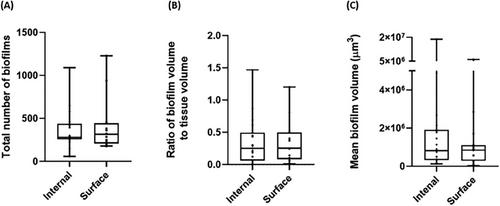
已知扁桃体中存在生物膜,但对其在扁桃体中的空间位置和大小分布却知之甚少。迄今为止,对扁桃体标本中生物膜的位置和分布的研究仅限于高倍率方法(如电子显微镜)或低倍率技术(如光学显微镜),前者能对微小的组织体积进行高分辨率成像,后者能对较大的标本进行成像,但空间分辨率较低。为了克服这些局限性,我们报告了使用多模态光学介镜观察和量化新鲜切除的小儿扁桃体中革兰氏阳性生物膜的数量和空间分布。这种方法可同时对扁桃体宿主和生物膜进行成像,成像范围可达 5 mm × 5 mm × 3 mm,整个成像具有亚细胞分辨率。对 36 个扁桃体标本进行的定量评估显示,扁桃体表面和扁桃体内部存在的生物膜在统计学上没有显著差异。这种新的定量中尺度成像方法可能有助于了解生物膜在扁桃体疾病和其他感染中的作用。
Multimodal optical mesoscopy reveals the quantity and spatial distribution of Gram-positive biofilms in ex vivo tonsils
Biofilms are known to be present in tonsils, but little is known about their spatial location and size distribution throughout the tonsil. Studies of the location and distribution of biofilms in tonsil specimens have thus far been limited to either high-magnification methods such as electron microscopy, which enables high-resolution imaging but only from a tiny tissue volume, or lower magnification techniques such as light microscopy, which allow imaging of larger specimens but with poor spatial resolution. To overcome these limitations, we report the use of multimodal optical mesoscopy to visualise and quantify the number and spatial distribution of Gram-positive biofilms in fresh, excised paediatric tonsils. This methodology supports simultaneous imaging of both the tonsil host and biofilms in whole mounts of tissue up to 5 mm × 5 mm × 3 mm with subcellular resolution throughout. A quantitative assessment of 36 tonsil specimens revealed no statistically significant difference between biofilm presence on the tonsil surface and the interior of the tonsil. This new quantitative mesoscale imaging approach may prove useful in understanding the role of biofilms in tonsillar diseases and other infections.
期刊介绍:
The Journal of Microscopy is the oldest journal dedicated to the science of microscopy and the only peer-reviewed publication of the Royal Microscopical Society. It publishes papers that report on the very latest developments in microscopy such as advances in microscopy techniques or novel areas of application. The Journal does not seek to publish routine applications of microscopy or specimen preparation even though the submission may otherwise have a high scientific merit.
The scope covers research in the physical and biological sciences and covers imaging methods using light, electrons, X-rays and other radiations as well as atomic force and near field techniques. Interdisciplinary research is welcome. Papers pertaining to microscopy are also welcomed on optical theory, spectroscopy, novel specimen preparation and manipulation methods and image recording, processing and analysis including dynamic analysis of living specimens.
Publication types include full papers, hot topic fast tracked communications and review articles. Authors considering submitting a review article should contact the editorial office first.


 分享
分享
 求助内容:
求助内容: 应助结果提醒方式:
 扫码关注我们
