Damiano Dei, Nicola Lambri, Leonardo Crespi, Ricardo Coimbra Brioso, Daniele Loiacono, Elena Clerici, Luisa Bellu, Chiara De Philippis, Pierina Navarria, Stefania Bramanti, Carmelo Carlo-Stella, Roberto Rusconi, Giacomo Reggiori, Stefano Tomatis, Marta Scorsetti, Pietro Mancosu
{"title":"基于深度学习和图集的模型,简化全骨髓和淋巴照射的分割工作流程","authors":"Damiano Dei, Nicola Lambri, Leonardo Crespi, Ricardo Coimbra Brioso, Daniele Loiacono, Elena Clerici, Luisa Bellu, Chiara De Philippis, Pierina Navarria, Stefania Bramanti, Carmelo Carlo-Stella, Roberto Rusconi, Giacomo Reggiori, Stefano Tomatis, Marta Scorsetti, Pietro Mancosu","doi":"10.1007/s11547-024-01760-8","DOIUrl":null,"url":null,"abstract":"<h3 data-test=\"abstract-sub-heading\">Purpose</h3><p>To improve the workflow of total marrow and lymphoid irradiation (TMLI) by enhancing the delineation of organs at risk (OARs) and clinical target volume (CTV) using deep learning (DL) and atlas-based (AB) segmentation models.</p><h3 data-test=\"abstract-sub-heading\">Materials and methods</h3><p>Ninety-five TMLI plans optimized in our institute were analyzed. Two commercial DL software were tested for segmenting 18 OARs. An AB model for lymph node CTV (CTV_LN) delineation was built using 20 TMLI patients. The AB model was evaluated on 20 independent patients, and a semiautomatic approach was tested by correcting the automatic contours. The generated OARs and CTV_LN contours were compared to manual contours in terms of topological agreement, dose statistics, and time workload. A clinical decision tree was developed to define a specific contouring strategy for each OAR.</p><h3 data-test=\"abstract-sub-heading\">Results</h3><p>The two DL models achieved a median [interquartile range] dice similarity coefficient (DSC) of 0.84 [0.71;0.93] and 0.85 [0.70;0.93] across the OARs. The absolute median Dmean difference between manual and the two DL models was 2.0 [0.7;6.6]% and 2.4 [0.9;7.1]%. The AB model achieved a median DSC of 0.70 [0.66;0.74] for CTV_LN delineation, increasing to 0.94 [0.94;0.95] after manual revision, with minimal Dmean differences. Since September 2022, our institution has implemented DL and AB models for all TMLI patients, reducing from 5 to 2 h the time required to complete the entire segmentation process.</p><h3 data-test=\"abstract-sub-heading\">Conclusion</h3><p>DL models can streamline the TMLI contouring process of OARs. Manual revision is still necessary for lymph node delineation using AB models.</p>","PeriodicalId":501689,"journal":{"name":"La radiologia medica","volume":"1 1","pages":""},"PeriodicalIF":0.0000,"publicationDate":"2024-02-02","publicationTypes":"Journal Article","fieldsOfStudy":null,"isOpenAccess":false,"openAccessPdf":"","citationCount":"0","resultStr":"{\"title\":\"Deep learning and atlas-based models to streamline the segmentation workflow of total marrow and lymphoid irradiation\",\"authors\":\"Damiano Dei, Nicola Lambri, Leonardo Crespi, Ricardo Coimbra Brioso, Daniele Loiacono, Elena Clerici, Luisa Bellu, Chiara De Philippis, Pierina Navarria, Stefania Bramanti, Carmelo Carlo-Stella, Roberto Rusconi, Giacomo Reggiori, Stefano Tomatis, Marta Scorsetti, Pietro Mancosu\",\"doi\":\"10.1007/s11547-024-01760-8\",\"DOIUrl\":null,\"url\":null,\"abstract\":\"<h3 data-test=\\\"abstract-sub-heading\\\">Purpose</h3><p>To improve the workflow of total marrow and lymphoid irradiation (TMLI) by enhancing the delineation of organs at risk (OARs) and clinical target volume (CTV) using deep learning (DL) and atlas-based (AB) segmentation models.</p><h3 data-test=\\\"abstract-sub-heading\\\">Materials and methods</h3><p>Ninety-five TMLI plans optimized in our institute were analyzed. Two commercial DL software were tested for segmenting 18 OARs. An AB model for lymph node CTV (CTV_LN) delineation was built using 20 TMLI patients. The AB model was evaluated on 20 independent patients, and a semiautomatic approach was tested by correcting the automatic contours. The generated OARs and CTV_LN contours were compared to manual contours in terms of topological agreement, dose statistics, and time workload. A clinical decision tree was developed to define a specific contouring strategy for each OAR.</p><h3 data-test=\\\"abstract-sub-heading\\\">Results</h3><p>The two DL models achieved a median [interquartile range] dice similarity coefficient (DSC) of 0.84 [0.71;0.93] and 0.85 [0.70;0.93] across the OARs. The absolute median Dmean difference between manual and the two DL models was 2.0 [0.7;6.6]% and 2.4 [0.9;7.1]%. The AB model achieved a median DSC of 0.70 [0.66;0.74] for CTV_LN delineation, increasing to 0.94 [0.94;0.95] after manual revision, with minimal Dmean differences. Since September 2022, our institution has implemented DL and AB models for all TMLI patients, reducing from 5 to 2 h the time required to complete the entire segmentation process.</p><h3 data-test=\\\"abstract-sub-heading\\\">Conclusion</h3><p>DL models can streamline the TMLI contouring process of OARs. Manual revision is still necessary for lymph node delineation using AB models.</p>\",\"PeriodicalId\":501689,\"journal\":{\"name\":\"La radiologia medica\",\"volume\":\"1 1\",\"pages\":\"\"},\"PeriodicalIF\":0.0000,\"publicationDate\":\"2024-02-02\",\"publicationTypes\":\"Journal Article\",\"fieldsOfStudy\":null,\"isOpenAccess\":false,\"openAccessPdf\":\"\",\"citationCount\":\"0\",\"resultStr\":null,\"platform\":\"Semanticscholar\",\"paperid\":null,\"PeriodicalName\":\"La radiologia medica\",\"FirstCategoryId\":\"1085\",\"ListUrlMain\":\"https://doi.org/10.1007/s11547-024-01760-8\",\"RegionNum\":0,\"RegionCategory\":null,\"ArticlePicture\":[],\"TitleCN\":null,\"AbstractTextCN\":null,\"PMCID\":null,\"EPubDate\":\"\",\"PubModel\":\"\",\"JCR\":\"\",\"JCRName\":\"\",\"Score\":null,\"Total\":0}","platform":"Semanticscholar","paperid":null,"PeriodicalName":"La radiologia medica","FirstCategoryId":"1085","ListUrlMain":"https://doi.org/10.1007/s11547-024-01760-8","RegionNum":0,"RegionCategory":null,"ArticlePicture":[],"TitleCN":null,"AbstractTextCN":null,"PMCID":null,"EPubDate":"","PubModel":"","JCR":"","JCRName":"","Score":null,"Total":0}
Deep learning and atlas-based models to streamline the segmentation workflow of total marrow and lymphoid irradiation
Purpose
To improve the workflow of total marrow and lymphoid irradiation (TMLI) by enhancing the delineation of organs at risk (OARs) and clinical target volume (CTV) using deep learning (DL) and atlas-based (AB) segmentation models.
Materials and methods
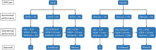
Ninety-five TMLI plans optimized in our institute were analyzed. Two commercial DL software were tested for segmenting 18 OARs. An AB model for lymph node CTV (CTV_LN) delineation was built using 20 TMLI patients. The AB model was evaluated on 20 independent patients, and a semiautomatic approach was tested by correcting the automatic contours. The generated OARs and CTV_LN contours were compared to manual contours in terms of topological agreement, dose statistics, and time workload. A clinical decision tree was developed to define a specific contouring strategy for each OAR.
Results
The two DL models achieved a median [interquartile range] dice similarity coefficient (DSC) of 0.84 [0.71;0.93] and 0.85 [0.70;0.93] across the OARs. The absolute median Dmean difference between manual and the two DL models was 2.0 [0.7;6.6]% and 2.4 [0.9;7.1]%. The AB model achieved a median DSC of 0.70 [0.66;0.74] for CTV_LN delineation, increasing to 0.94 [0.94;0.95] after manual revision, with minimal Dmean differences. Since September 2022, our institution has implemented DL and AB models for all TMLI patients, reducing from 5 to 2 h the time required to complete the entire segmentation process.
Conclusion
DL models can streamline the TMLI contouring process of OARs. Manual revision is still necessary for lymph node delineation using AB models.


 分享
分享

 求助内容:
求助内容: 应助结果提醒方式:
 扫码关注我们
