Xiwei Fan, Antonia Rujia Sun, Reuben S. E. Young, Isaac O. Afara, Brett R. Hamilton, Louis Jun Ye Ong, Ross Crawford, Indira Prasadam
{"title":"骨关节炎微环境的空间分析:技术、见解和应用","authors":"Xiwei Fan, Antonia Rujia Sun, Reuben S. E. Young, Isaac O. Afara, Brett R. Hamilton, Louis Jun Ye Ong, Ross Crawford, Indira Prasadam","doi":"10.1038/s41413-023-00304-6","DOIUrl":null,"url":null,"abstract":"<p>Osteoarthritis (OA) is a debilitating degenerative disease affecting multiple joint tissues, including cartilage, bone, synovium, and adipose tissues. OA presents diverse clinical phenotypes and distinct molecular endotypes, including inflammatory, metabolic, mechanical, genetic, and synovial variants. Consequently, innovative technologies are needed to support the development of effective diagnostic and precision therapeutic approaches. Traditional analysis of bulk OA tissue extracts has limitations due to technical constraints, causing challenges in the differentiation between various physiological and pathological phenotypes in joint tissues. This issue has led to standardization difficulties and hindered the success of clinical trials. Gaining insights into the spatial variations of the cellular and molecular structures in OA tissues, encompassing DNA, RNA, metabolites, and proteins, as well as their chemical properties, elemental composition, and mechanical attributes, can contribute to a more comprehensive understanding of the disease subtypes. Spatially resolved biology enables biologists to investigate cells within the context of their tissue microenvironment, providing a more holistic view of cellular function. Recent advances in innovative spatial biology techniques now allow intact tissue sections to be examined using various -omics lenses, such as genomics, transcriptomics, proteomics, and metabolomics, with spatial data. This fusion of approaches provides researchers with critical insights into the molecular composition and functions of the cells and tissues at precise spatial coordinates. Furthermore, advanced imaging techniques, including high-resolution microscopy, hyperspectral imaging, and mass spectrometry imaging, enable the visualization and analysis of the spatial distribution of biomolecules, cells, and tissues. Linking these molecular imaging outputs to conventional tissue histology can facilitate a more comprehensive characterization of disease phenotypes. This review summarizes the recent advancements in the molecular imaging modalities and methodologies for in-depth spatial analysis. It explores their applications, challenges, and potential opportunities in the field of OA. Additionally, this review provides a perspective on the potential research directions for these contemporary approaches that can meet the requirements of clinical diagnoses and the establishment of therapeutic targets for OA.</p>","PeriodicalId":9134,"journal":{"name":"Bone Research","volume":"30 1","pages":""},"PeriodicalIF":15.0000,"publicationDate":"2024-02-04","publicationTypes":"Journal Article","fieldsOfStudy":null,"isOpenAccess":false,"openAccessPdf":"","citationCount":"0","resultStr":"{\"title\":\"Spatial analysis of the osteoarthritis microenvironment: techniques, insights, and applications\",\"authors\":\"Xiwei Fan, Antonia Rujia Sun, Reuben S. E. Young, Isaac O. Afara, Brett R. Hamilton, Louis Jun Ye Ong, Ross Crawford, Indira Prasadam\",\"doi\":\"10.1038/s41413-023-00304-6\",\"DOIUrl\":null,\"url\":null,\"abstract\":\"<p>Osteoarthritis (OA) is a debilitating degenerative disease affecting multiple joint tissues, including cartilage, bone, synovium, and adipose tissues. OA presents diverse clinical phenotypes and distinct molecular endotypes, including inflammatory, metabolic, mechanical, genetic, and synovial variants. Consequently, innovative technologies are needed to support the development of effective diagnostic and precision therapeutic approaches. Traditional analysis of bulk OA tissue extracts has limitations due to technical constraints, causing challenges in the differentiation between various physiological and pathological phenotypes in joint tissues. This issue has led to standardization difficulties and hindered the success of clinical trials. Gaining insights into the spatial variations of the cellular and molecular structures in OA tissues, encompassing DNA, RNA, metabolites, and proteins, as well as their chemical properties, elemental composition, and mechanical attributes, can contribute to a more comprehensive understanding of the disease subtypes. Spatially resolved biology enables biologists to investigate cells within the context of their tissue microenvironment, providing a more holistic view of cellular function. Recent advances in innovative spatial biology techniques now allow intact tissue sections to be examined using various -omics lenses, such as genomics, transcriptomics, proteomics, and metabolomics, with spatial data. This fusion of approaches provides researchers with critical insights into the molecular composition and functions of the cells and tissues at precise spatial coordinates. Furthermore, advanced imaging techniques, including high-resolution microscopy, hyperspectral imaging, and mass spectrometry imaging, enable the visualization and analysis of the spatial distribution of biomolecules, cells, and tissues. Linking these molecular imaging outputs to conventional tissue histology can facilitate a more comprehensive characterization of disease phenotypes. This review summarizes the recent advancements in the molecular imaging modalities and methodologies for in-depth spatial analysis. It explores their applications, challenges, and potential opportunities in the field of OA. Additionally, this review provides a perspective on the potential research directions for these contemporary approaches that can meet the requirements of clinical diagnoses and the establishment of therapeutic targets for OA.</p>\",\"PeriodicalId\":9134,\"journal\":{\"name\":\"Bone Research\",\"volume\":\"30 1\",\"pages\":\"\"},\"PeriodicalIF\":15.0000,\"publicationDate\":\"2024-02-04\",\"publicationTypes\":\"Journal Article\",\"fieldsOfStudy\":null,\"isOpenAccess\":false,\"openAccessPdf\":\"\",\"citationCount\":\"0\",\"resultStr\":null,\"platform\":\"Semanticscholar\",\"paperid\":null,\"PeriodicalName\":\"Bone Research\",\"FirstCategoryId\":\"3\",\"ListUrlMain\":\"https://doi.org/10.1038/s41413-023-00304-6\",\"RegionNum\":1,\"RegionCategory\":\"医学\",\"ArticlePicture\":[],\"TitleCN\":null,\"AbstractTextCN\":null,\"PMCID\":null,\"EPubDate\":\"\",\"PubModel\":\"\",\"JCR\":\"Q1\",\"JCRName\":\"CELL & TISSUE ENGINEERING\",\"Score\":null,\"Total\":0}","platform":"Semanticscholar","paperid":null,"PeriodicalName":"Bone Research","FirstCategoryId":"3","ListUrlMain":"https://doi.org/10.1038/s41413-023-00304-6","RegionNum":1,"RegionCategory":"医学","ArticlePicture":[],"TitleCN":null,"AbstractTextCN":null,"PMCID":null,"EPubDate":"","PubModel":"","JCR":"Q1","JCRName":"CELL & TISSUE ENGINEERING","Score":null,"Total":0}
引用次数: 0
摘要
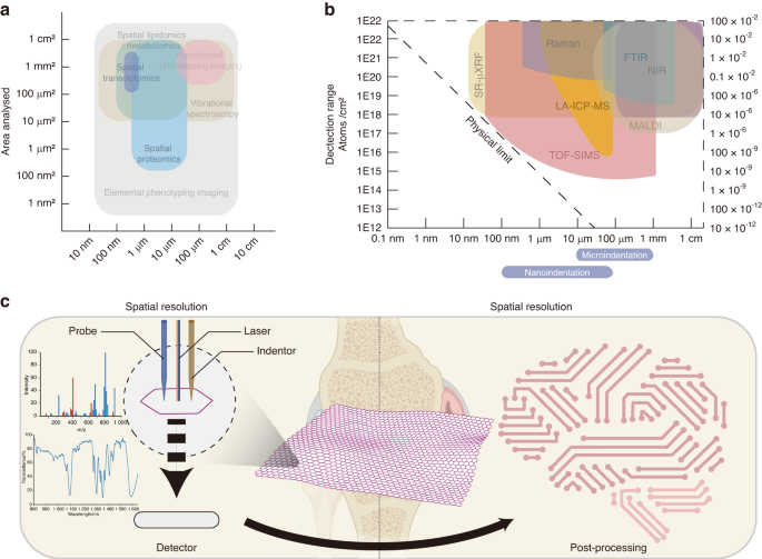
骨关节炎(OA)是一种使人衰弱的退行性疾病,影响多个关节组织,包括软骨、骨、滑膜和脂肪组织。OA 表现出多种临床表型和不同的分子内型,包括炎症、代谢、机械、遗传和滑膜变异。因此,需要创新技术来支持有效诊断和精准治疗方法的开发。由于技术限制,对大块 OA 组织提取物的传统分析存在局限性,给区分关节组织中的各种生理和病理表型带来了挑战。这一问题导致标准化困难,阻碍了临床试验的成功。深入了解 OA 组织中细胞和分子结构的空间变化,包括 DNA、RNA、代谢物和蛋白质,以及它们的化学特性、元素组成和机械属性,有助于更全面地了解疾病亚型。空间解析生物学使生物学家能够在细胞组织微环境的背景下研究细胞,从而更全面地了解细胞功能。创新性空间生物学技术的最新进展使完整的组织切片现在可以利用基因组学、转录组学、蛋白质组学和代谢组学等各种组学透镜和空间数据进行研究。这种方法的融合为研究人员在精确的空间坐标上深入了解细胞和组织的分子组成和功能提供了重要依据。此外,先进的成像技术,包括高分辨率显微镜、高光谱成像和质谱成像,使生物分子、细胞和组织空间分布的可视化和分析成为可能。将这些分子成像结果与传统的组织组织学联系起来,有助于更全面地描述疾病表型。本综述总结了分子成像模式和深入空间分析方法的最新进展。它探讨了它们在 OA 领域的应用、挑战和潜在机遇。此外,本综述还对这些当代方法的潜在研究方向提供了一个视角,以满足临床诊断和确立 OA 治疗靶点的要求。
Spatial analysis of the osteoarthritis microenvironment: techniques, insights, and applications
Osteoarthritis (OA) is a debilitating degenerative disease affecting multiple joint tissues, including cartilage, bone, synovium, and adipose tissues. OA presents diverse clinical phenotypes and distinct molecular endotypes, including inflammatory, metabolic, mechanical, genetic, and synovial variants. Consequently, innovative technologies are needed to support the development of effective diagnostic and precision therapeutic approaches. Traditional analysis of bulk OA tissue extracts has limitations due to technical constraints, causing challenges in the differentiation between various physiological and pathological phenotypes in joint tissues. This issue has led to standardization difficulties and hindered the success of clinical trials. Gaining insights into the spatial variations of the cellular and molecular structures in OA tissues, encompassing DNA, RNA, metabolites, and proteins, as well as their chemical properties, elemental composition, and mechanical attributes, can contribute to a more comprehensive understanding of the disease subtypes. Spatially resolved biology enables biologists to investigate cells within the context of their tissue microenvironment, providing a more holistic view of cellular function. Recent advances in innovative spatial biology techniques now allow intact tissue sections to be examined using various -omics lenses, such as genomics, transcriptomics, proteomics, and metabolomics, with spatial data. This fusion of approaches provides researchers with critical insights into the molecular composition and functions of the cells and tissues at precise spatial coordinates. Furthermore, advanced imaging techniques, including high-resolution microscopy, hyperspectral imaging, and mass spectrometry imaging, enable the visualization and analysis of the spatial distribution of biomolecules, cells, and tissues. Linking these molecular imaging outputs to conventional tissue histology can facilitate a more comprehensive characterization of disease phenotypes. This review summarizes the recent advancements in the molecular imaging modalities and methodologies for in-depth spatial analysis. It explores their applications, challenges, and potential opportunities in the field of OA. Additionally, this review provides a perspective on the potential research directions for these contemporary approaches that can meet the requirements of clinical diagnoses and the establishment of therapeutic targets for OA.
期刊介绍:
Established in 2013, Bone Research is a newly-founded English-language periodical that centers on the basic and clinical facets of bone biology, pathophysiology, and regeneration. It is dedicated to championing key findings emerging from both basic investigations and clinical research concerning bone-related topics. The journal's objective is to globally disseminate research in bone-related physiology, pathology, diseases, and treatment, contributing to the advancement of knowledge in this field.


 分享
分享
 求助内容:
求助内容: 应助结果提醒方式:
 扫码关注我们
