Sung Hyun Kang, Hyeon Jin Park, Jae-Won Hyun, Ho-Shin Gwak
{"title":"一名儿童脑胶质瘤模拟脑干病变的自发消退:病例报告。","authors":"Sung Hyun Kang, Hyeon Jin Park, Jae-Won Hyun, Ho-Shin Gwak","doi":"10.14791/btrt.2023.0039","DOIUrl":null,"url":null,"abstract":"<p><p>Differential diagnosis of focal brainstem lesions detected on MRI is challenging, especially in young children. Formerly, brainstem gliomas were classified mainly based on MRI features and location. However, since 2016, the World Health Organization's brainstem lesion classification requires tissue biopsy to reveal molecular characteristics. Although modern techniques of stereotactic or navigation-guided biopsy ensure accurate biopsy of the lesion with safety, biopsy of brainstem lesions is still generally not performed. Here, we report a focal brainstem lesion mimicking brainstem glioma in a 9-year-old girl. Initial MRI, MR spectroscopy, and <sup>11</sup>C-methionine positron emission tomography (PET) features suggested low-grade glioma or diffuse intrinsic pontine glioma. However, repeated MR spectroscopy, perfusion MRI, and <sup>18</sup>fluorodeoxyglucose PET findings suggested that it was more likely a non-tumorous lesion. As the patient presented not with a neurological manifestation but with precocious puberty, the attending oncologist chose to observe with regular follow-up MRI. The pontine lesion with high signal intensity on T2-weighted MRI regressed from the 6-month follow-up and became invisible on the 1.5-year follow-up MRI. We reviewed brainstem glioma-mimicking lesions in the literature and discussed the key points of differential diagnosis.</p>","PeriodicalId":72453,"journal":{"name":"Brain tumor research and treatment","volume":"12 1","pages":"58-62"},"PeriodicalIF":0.0000,"publicationDate":"2024-01-01","publicationTypes":"Journal Article","fieldsOfStudy":null,"isOpenAccess":false,"openAccessPdf":"https://www.ncbi.nlm.nih.gov/pmc/articles/PMC10864136/pdf/","citationCount":"0","resultStr":"{\"title\":\"Spontaneous Regression of Glioma-Mimicking Brainstem Lesion in a Child: A Case Report.\",\"authors\":\"Sung Hyun Kang, Hyeon Jin Park, Jae-Won Hyun, Ho-Shin Gwak\",\"doi\":\"10.14791/btrt.2023.0039\",\"DOIUrl\":null,\"url\":null,\"abstract\":\"<p><p>Differential diagnosis of focal brainstem lesions detected on MRI is challenging, especially in young children. Formerly, brainstem gliomas were classified mainly based on MRI features and location. However, since 2016, the World Health Organization's brainstem lesion classification requires tissue biopsy to reveal molecular characteristics. Although modern techniques of stereotactic or navigation-guided biopsy ensure accurate biopsy of the lesion with safety, biopsy of brainstem lesions is still generally not performed. Here, we report a focal brainstem lesion mimicking brainstem glioma in a 9-year-old girl. Initial MRI, MR spectroscopy, and <sup>11</sup>C-methionine positron emission tomography (PET) features suggested low-grade glioma or diffuse intrinsic pontine glioma. However, repeated MR spectroscopy, perfusion MRI, and <sup>18</sup>fluorodeoxyglucose PET findings suggested that it was more likely a non-tumorous lesion. As the patient presented not with a neurological manifestation but with precocious puberty, the attending oncologist chose to observe with regular follow-up MRI. The pontine lesion with high signal intensity on T2-weighted MRI regressed from the 6-month follow-up and became invisible on the 1.5-year follow-up MRI. We reviewed brainstem glioma-mimicking lesions in the literature and discussed the key points of differential diagnosis.</p>\",\"PeriodicalId\":72453,\"journal\":{\"name\":\"Brain tumor research and treatment\",\"volume\":\"12 1\",\"pages\":\"58-62\"},\"PeriodicalIF\":0.0000,\"publicationDate\":\"2024-01-01\",\"publicationTypes\":\"Journal Article\",\"fieldsOfStudy\":null,\"isOpenAccess\":false,\"openAccessPdf\":\"https://www.ncbi.nlm.nih.gov/pmc/articles/PMC10864136/pdf/\",\"citationCount\":\"0\",\"resultStr\":null,\"platform\":\"Semanticscholar\",\"paperid\":null,\"PeriodicalName\":\"Brain tumor research and treatment\",\"FirstCategoryId\":\"1085\",\"ListUrlMain\":\"https://doi.org/10.14791/btrt.2023.0039\",\"RegionNum\":0,\"RegionCategory\":null,\"ArticlePicture\":[],\"TitleCN\":null,\"AbstractTextCN\":null,\"PMCID\":null,\"EPubDate\":\"\",\"PubModel\":\"\",\"JCR\":\"\",\"JCRName\":\"\",\"Score\":null,\"Total\":0}","platform":"Semanticscholar","paperid":null,"PeriodicalName":"Brain tumor research and treatment","FirstCategoryId":"1085","ListUrlMain":"https://doi.org/10.14791/btrt.2023.0039","RegionNum":0,"RegionCategory":null,"ArticlePicture":[],"TitleCN":null,"AbstractTextCN":null,"PMCID":null,"EPubDate":"","PubModel":"","JCR":"","JCRName":"","Score":null,"Total":0}
Spontaneous Regression of Glioma-Mimicking Brainstem Lesion in a Child: A Case Report.
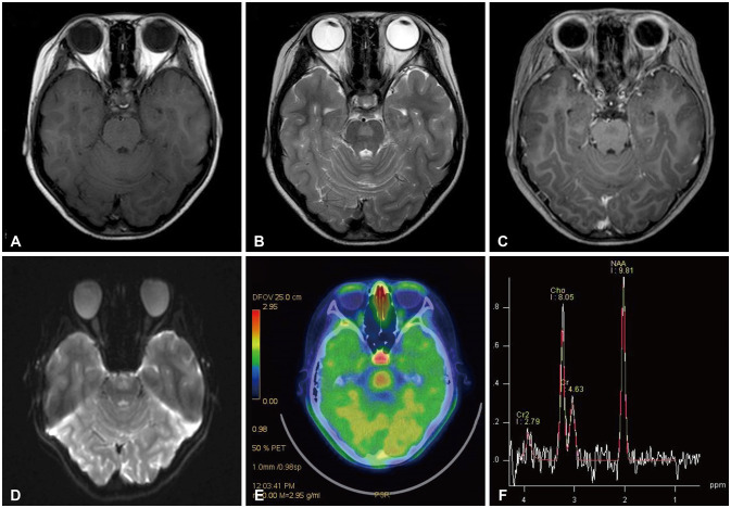
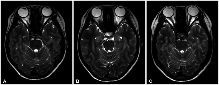
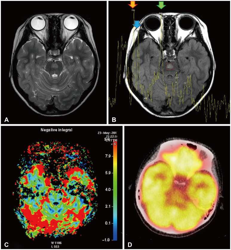
Differential diagnosis of focal brainstem lesions detected on MRI is challenging, especially in young children. Formerly, brainstem gliomas were classified mainly based on MRI features and location. However, since 2016, the World Health Organization's brainstem lesion classification requires tissue biopsy to reveal molecular characteristics. Although modern techniques of stereotactic or navigation-guided biopsy ensure accurate biopsy of the lesion with safety, biopsy of brainstem lesions is still generally not performed. Here, we report a focal brainstem lesion mimicking brainstem glioma in a 9-year-old girl. Initial MRI, MR spectroscopy, and 11C-methionine positron emission tomography (PET) features suggested low-grade glioma or diffuse intrinsic pontine glioma. However, repeated MR spectroscopy, perfusion MRI, and 18fluorodeoxyglucose PET findings suggested that it was more likely a non-tumorous lesion. As the patient presented not with a neurological manifestation but with precocious puberty, the attending oncologist chose to observe with regular follow-up MRI. The pontine lesion with high signal intensity on T2-weighted MRI regressed from the 6-month follow-up and became invisible on the 1.5-year follow-up MRI. We reviewed brainstem glioma-mimicking lesions in the literature and discussed the key points of differential diagnosis.


 分享
分享

 求助内容:
求助内容: 应助结果提醒方式:
 扫码关注我们
