Ian MacLaren, Enrique Frutos-Myro, Steven Zeltmann, Colin Ophus
{"title":"利用扫描前驱电子衍射和开源软件库,以纳米分辨率绘制α-β钛合金晶体图的方法。","authors":"Ian MacLaren, Enrique Frutos-Myro, Steven Zeltmann, Colin Ophus","doi":"10.1111/jmi.13275","DOIUrl":null,"url":null,"abstract":"<p>An approach for the crystallographic mapping of two-phase alloys on the nanoscale using a combination of scanned precession electron diffraction and open-source python libraries is introduced in this paper. This method is demonstrated using the example of a two-phase α/β titanium alloy. The data were recorded using a direct electron detector to collect the patterns, and recently developed algorithms to perform automated indexing and analyse the crystallography from the results. Very high-quality mapping is achieved at a 3 nm step size. The results show the expected Burgers orientation relationships between the α laths and β matrix, as well as the expected misorientations between α laths. A minor issue was found that one area was affected by 180° ambiguities in indexing occur due to this area being aligned too close to a zone axis of the α with twofold projection symmetry (not present in 3D) in the zero-order Laue Zone, and this should be avoided in data acquisition in the future. Nevertheless, this study demonstrates a good workflow for the analysis of nanocrystalline two- or multi-phase materials, which will be of widespread use in analysing two-phase titanium and other systems and how they evolve as a function of thermomechanical treatments.</p>","PeriodicalId":16484,"journal":{"name":"Journal of microscopy","volume":"295 2","pages":"131-139"},"PeriodicalIF":1.9000,"publicationDate":"2024-02-14","publicationTypes":"Journal Article","fieldsOfStudy":null,"isOpenAccess":false,"openAccessPdf":"https://onlinelibrary.wiley.com/doi/epdf/10.1111/jmi.13275","citationCount":"0","resultStr":"{\"title\":\"A method for crystallographic mapping of an alpha-beta titanium alloy with nanometre resolution using scanning precession electron diffraction and open-source software libraries\",\"authors\":\"Ian MacLaren, Enrique Frutos-Myro, Steven Zeltmann, Colin Ophus\",\"doi\":\"10.1111/jmi.13275\",\"DOIUrl\":null,\"url\":null,\"abstract\":\"<p>An approach for the crystallographic mapping of two-phase alloys on the nanoscale using a combination of scanned precession electron diffraction and open-source python libraries is introduced in this paper. This method is demonstrated using the example of a two-phase α/β titanium alloy. The data were recorded using a direct electron detector to collect the patterns, and recently developed algorithms to perform automated indexing and analyse the crystallography from the results. Very high-quality mapping is achieved at a 3 nm step size. The results show the expected Burgers orientation relationships between the α laths and β matrix, as well as the expected misorientations between α laths. A minor issue was found that one area was affected by 180° ambiguities in indexing occur due to this area being aligned too close to a zone axis of the α with twofold projection symmetry (not present in 3D) in the zero-order Laue Zone, and this should be avoided in data acquisition in the future. Nevertheless, this study demonstrates a good workflow for the analysis of nanocrystalline two- or multi-phase materials, which will be of widespread use in analysing two-phase titanium and other systems and how they evolve as a function of thermomechanical treatments.</p>\",\"PeriodicalId\":16484,\"journal\":{\"name\":\"Journal of microscopy\",\"volume\":\"295 2\",\"pages\":\"131-139\"},\"PeriodicalIF\":1.9000,\"publicationDate\":\"2024-02-14\",\"publicationTypes\":\"Journal Article\",\"fieldsOfStudy\":null,\"isOpenAccess\":false,\"openAccessPdf\":\"https://onlinelibrary.wiley.com/doi/epdf/10.1111/jmi.13275\",\"citationCount\":\"0\",\"resultStr\":null,\"platform\":\"Semanticscholar\",\"paperid\":null,\"PeriodicalName\":\"Journal of microscopy\",\"FirstCategoryId\":\"5\",\"ListUrlMain\":\"https://onlinelibrary.wiley.com/doi/10.1111/jmi.13275\",\"RegionNum\":4,\"RegionCategory\":\"工程技术\",\"ArticlePicture\":[],\"TitleCN\":null,\"AbstractTextCN\":null,\"PMCID\":null,\"EPubDate\":\"\",\"PubModel\":\"\",\"JCR\":\"Q3\",\"JCRName\":\"MICROSCOPY\",\"Score\":null,\"Total\":0}","platform":"Semanticscholar","paperid":null,"PeriodicalName":"Journal of microscopy","FirstCategoryId":"5","ListUrlMain":"https://onlinelibrary.wiley.com/doi/10.1111/jmi.13275","RegionNum":4,"RegionCategory":"工程技术","ArticlePicture":[],"TitleCN":null,"AbstractTextCN":null,"PMCID":null,"EPubDate":"","PubModel":"","JCR":"Q3","JCRName":"MICROSCOPY","Score":null,"Total":0}
A method for crystallographic mapping of an alpha-beta titanium alloy with nanometre resolution using scanning precession electron diffraction and open-source software libraries
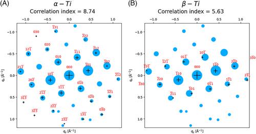
An approach for the crystallographic mapping of two-phase alloys on the nanoscale using a combination of scanned precession electron diffraction and open-source python libraries is introduced in this paper. This method is demonstrated using the example of a two-phase α/β titanium alloy. The data were recorded using a direct electron detector to collect the patterns, and recently developed algorithms to perform automated indexing and analyse the crystallography from the results. Very high-quality mapping is achieved at a 3 nm step size. The results show the expected Burgers orientation relationships between the α laths and β matrix, as well as the expected misorientations between α laths. A minor issue was found that one area was affected by 180° ambiguities in indexing occur due to this area being aligned too close to a zone axis of the α with twofold projection symmetry (not present in 3D) in the zero-order Laue Zone, and this should be avoided in data acquisition in the future. Nevertheless, this study demonstrates a good workflow for the analysis of nanocrystalline two- or multi-phase materials, which will be of widespread use in analysing two-phase titanium and other systems and how they evolve as a function of thermomechanical treatments.
期刊介绍:
The Journal of Microscopy is the oldest journal dedicated to the science of microscopy and the only peer-reviewed publication of the Royal Microscopical Society. It publishes papers that report on the very latest developments in microscopy such as advances in microscopy techniques or novel areas of application. The Journal does not seek to publish routine applications of microscopy or specimen preparation even though the submission may otherwise have a high scientific merit.
The scope covers research in the physical and biological sciences and covers imaging methods using light, electrons, X-rays and other radiations as well as atomic force and near field techniques. Interdisciplinary research is welcome. Papers pertaining to microscopy are also welcomed on optical theory, spectroscopy, novel specimen preparation and manipulation methods and image recording, processing and analysis including dynamic analysis of living specimens.
Publication types include full papers, hot topic fast tracked communications and review articles. Authors considering submitting a review article should contact the editorial office first.


 分享
分享
 求助内容:
求助内容: 应助结果提醒方式:
 扫码关注我们
