Hong Wu, Yoshihiko Fujioka, Shoichi Sakaguchi, Youichi Suzuki, Takashi Nakano
{"title":"对 SARS-CoV-2 感染细胞空泡中的两种病毒颗粒进行形态学分析。","authors":"Hong Wu, Yoshihiko Fujioka, Shoichi Sakaguchi, Youichi Suzuki, Takashi Nakano","doi":"10.1007/s00795-024-00381-4","DOIUrl":null,"url":null,"abstract":"<p><p>In this study, we analyzed the morphological structure of severe acute respiratory syndrome coronavirus 2 (SARS-CoV-2) in human cells. We identified the two types of viral particles present within the vacuoles of infected cells. Using transmission electron microscopy, we observed that SARS-CoV-2 particles exhibited both low- and high-electron-density structures, which was further confirmed through three-dimensional reconstruction using electron tomography. The budding of these particles was exclusively observed within these vacuoles. Intriguingly, viral particles with low-electron-density structures were confined to vacuoles, whereas those with high-electron-density structures were found in vacuoles and on the cell membrane surface of infected cells. Notably, high-electron-density particles found within vacuoles exhibited the same morphology as those outside the infected cells. This observation suggests that the two types of viral particles identified in this study had different maturation status. Our findings provide valuable insights into the molecular details of SARS-CoV-2 particles, contributing to our understanding of the virus.</p>","PeriodicalId":18338,"journal":{"name":"Medical Molecular Morphology","volume":" ","pages":"124-135"},"PeriodicalIF":1.1000,"publicationDate":"2024-06-01","publicationTypes":"Journal Article","fieldsOfStudy":null,"isOpenAccess":false,"openAccessPdf":"","citationCount":"0","resultStr":"{\"title\":\"Morphological analysis for two types of viral particles in vacuoles of SARS-CoV-2-infected cells.\",\"authors\":\"Hong Wu, Yoshihiko Fujioka, Shoichi Sakaguchi, Youichi Suzuki, Takashi Nakano\",\"doi\":\"10.1007/s00795-024-00381-4\",\"DOIUrl\":null,\"url\":null,\"abstract\":\"<p><p>In this study, we analyzed the morphological structure of severe acute respiratory syndrome coronavirus 2 (SARS-CoV-2) in human cells. We identified the two types of viral particles present within the vacuoles of infected cells. Using transmission electron microscopy, we observed that SARS-CoV-2 particles exhibited both low- and high-electron-density structures, which was further confirmed through three-dimensional reconstruction using electron tomography. The budding of these particles was exclusively observed within these vacuoles. Intriguingly, viral particles with low-electron-density structures were confined to vacuoles, whereas those with high-electron-density structures were found in vacuoles and on the cell membrane surface of infected cells. Notably, high-electron-density particles found within vacuoles exhibited the same morphology as those outside the infected cells. This observation suggests that the two types of viral particles identified in this study had different maturation status. Our findings provide valuable insights into the molecular details of SARS-CoV-2 particles, contributing to our understanding of the virus.</p>\",\"PeriodicalId\":18338,\"journal\":{\"name\":\"Medical Molecular Morphology\",\"volume\":\" \",\"pages\":\"124-135\"},\"PeriodicalIF\":1.1000,\"publicationDate\":\"2024-06-01\",\"publicationTypes\":\"Journal Article\",\"fieldsOfStudy\":null,\"isOpenAccess\":false,\"openAccessPdf\":\"\",\"citationCount\":\"0\",\"resultStr\":null,\"platform\":\"Semanticscholar\",\"paperid\":null,\"PeriodicalName\":\"Medical Molecular Morphology\",\"FirstCategoryId\":\"3\",\"ListUrlMain\":\"https://doi.org/10.1007/s00795-024-00381-4\",\"RegionNum\":4,\"RegionCategory\":\"医学\",\"ArticlePicture\":[],\"TitleCN\":null,\"AbstractTextCN\":null,\"PMCID\":null,\"EPubDate\":\"2024/2/23 0:00:00\",\"PubModel\":\"Epub\",\"JCR\":\"Q3\",\"JCRName\":\"PATHOLOGY\",\"Score\":null,\"Total\":0}","platform":"Semanticscholar","paperid":null,"PeriodicalName":"Medical Molecular Morphology","FirstCategoryId":"3","ListUrlMain":"https://doi.org/10.1007/s00795-024-00381-4","RegionNum":4,"RegionCategory":"医学","ArticlePicture":[],"TitleCN":null,"AbstractTextCN":null,"PMCID":null,"EPubDate":"2024/2/23 0:00:00","PubModel":"Epub","JCR":"Q3","JCRName":"PATHOLOGY","Score":null,"Total":0}
Morphological analysis for two types of viral particles in vacuoles of SARS-CoV-2-infected cells.
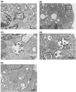
In this study, we analyzed the morphological structure of severe acute respiratory syndrome coronavirus 2 (SARS-CoV-2) in human cells. We identified the two types of viral particles present within the vacuoles of infected cells. Using transmission electron microscopy, we observed that SARS-CoV-2 particles exhibited both low- and high-electron-density structures, which was further confirmed through three-dimensional reconstruction using electron tomography. The budding of these particles was exclusively observed within these vacuoles. Intriguingly, viral particles with low-electron-density structures were confined to vacuoles, whereas those with high-electron-density structures were found in vacuoles and on the cell membrane surface of infected cells. Notably, high-electron-density particles found within vacuoles exhibited the same morphology as those outside the infected cells. This observation suggests that the two types of viral particles identified in this study had different maturation status. Our findings provide valuable insights into the molecular details of SARS-CoV-2 particles, contributing to our understanding of the virus.
期刊介绍:
Medical Molecular Morphology is an international forum for researchers in both basic and clinical medicine to present and discuss new research on the structural mechanisms and the processes of health and disease at the molecular level. The structures of molecules, organelles, cells, tissues, and organs determine their normal function. Disease is thus best understood in terms of structural changes in these different levels of biological organization, especially in molecules and molecular interactions as well as the cellular localization of chemical components. Medical Molecular Morphology welcomes articles on basic or clinical research in the fields of cell biology, molecular biology, and medical, veterinary, and dental sciences using techniques for structural research such as electron microscopy, confocal laser scanning microscopy, enzyme histochemistry, immunohistochemistry, radioautography, X-ray microanalysis, and in situ hybridization.
Manuscripts submitted for publication must contain a statement to the effect that all human studies have been reviewed by the appropriate ethics committee and have therefore been performed in accordance with the ethical standards laid down in an appropriate version of the 1964 Declaration of Helsinki. It should also be stated clearly in the text that all persons gave their informed consent prior to their inclusion in the study. Details that might disclose the identity of the subjects under study should be omitted.


 分享
分享
 求助内容:
求助内容: 应助结果提醒方式:
 扫码关注我们
