{"title":"用免疫组化方法诊断犬纺锤形细胞瘤的挑战,以三例来自裸眼膜的非色素性恶性病例为例。","authors":"Kristine Bundgaard Kjellingbro, Carolina Naranjo Freixa, Lauge Hjorth Mikkelsen, Steffen Heegaard","doi":"10.1186/s13028-024-00727-z","DOIUrl":null,"url":null,"abstract":"<p><strong>Background: </strong>Nonpigmented malignant spindle cell tumours of the membrana nictitans are rare in dogs. In twenty-three years only three cases have been diagnosed in Scandinavia. This study describes the three cases of malignant tumours of the membrana nictitans recorded by the Eye Pathology Section, University of Copenhagen, Denmark, with reference to the clinical appearance and work-up, the treatment and prognosis, and the histopathological description including immunohistochemistry. The three cases are compared to previous publications on canine tumours of the nictitating membrane. We emphasize the importance of using protocols that are adapted to the specific species such as dogs. Opposite the human tissue responses, we even need more than one marker when diagnosing melanomas in dogs.</p><p><strong>Results: </strong>The dogs presented were an 8-year-old Dachshund, a 12-year-old Akita and a 14-year-old Shetland Sheepdog. All three dogs were entire females. All three nictitating membrane tumours developed on the right nictitating membrane as firm or multilobulated hyperaemic masses. Two of the tumours were macroscopically nonpigmented, the third being partly pigmented on the surface and ulcerated. According to the histopathology and for two of the cases immunohistochemistry with dog-adapted protocols the diagnoses included one hemangiosarcoma and two amelanotic melanomas. Tumour regrowth developed in all three cases and repeated resections were completed 1, 2 and 3 times, respectively, with recurrence experienced within 1.5 months - 3 years.</p><p><strong>Conclusions: </strong>Nonpigmented malignant spindle cell tumours of the canine membrana nictitans are rare. Treatment of choice should be complete excision with a minimal histologic tumour-free distance and in case of a recurrence a full resection of the nictitating membrane. We strongly recommend a dog-adapted protocol for immunohistochemistry.</p>","PeriodicalId":7181,"journal":{"name":"Acta Veterinaria Scandinavica","volume":"66 1","pages":"7"},"PeriodicalIF":1.7000,"publicationDate":"2024-02-23","publicationTypes":"Journal Article","fieldsOfStudy":null,"isOpenAccess":false,"openAccessPdf":"https://www.ncbi.nlm.nih.gov/pmc/articles/PMC10893616/pdf/","citationCount":"0","resultStr":"{\"title\":\"Challenges in diagnosing canine spindle cell tumours using immunohistochemistry, illustrated by three nonpigmented malignant cases from the nictitating membrane.\",\"authors\":\"Kristine Bundgaard Kjellingbro, Carolina Naranjo Freixa, Lauge Hjorth Mikkelsen, Steffen Heegaard\",\"doi\":\"10.1186/s13028-024-00727-z\",\"DOIUrl\":null,\"url\":null,\"abstract\":\"<p><strong>Background: </strong>Nonpigmented malignant spindle cell tumours of the membrana nictitans are rare in dogs. In twenty-three years only three cases have been diagnosed in Scandinavia. This study describes the three cases of malignant tumours of the membrana nictitans recorded by the Eye Pathology Section, University of Copenhagen, Denmark, with reference to the clinical appearance and work-up, the treatment and prognosis, and the histopathological description including immunohistochemistry. The three cases are compared to previous publications on canine tumours of the nictitating membrane. We emphasize the importance of using protocols that are adapted to the specific species such as dogs. Opposite the human tissue responses, we even need more than one marker when diagnosing melanomas in dogs.</p><p><strong>Results: </strong>The dogs presented were an 8-year-old Dachshund, a 12-year-old Akita and a 14-year-old Shetland Sheepdog. All three dogs were entire females. All three nictitating membrane tumours developed on the right nictitating membrane as firm or multilobulated hyperaemic masses. Two of the tumours were macroscopically nonpigmented, the third being partly pigmented on the surface and ulcerated. According to the histopathology and for two of the cases immunohistochemistry with dog-adapted protocols the diagnoses included one hemangiosarcoma and two amelanotic melanomas. Tumour regrowth developed in all three cases and repeated resections were completed 1, 2 and 3 times, respectively, with recurrence experienced within 1.5 months - 3 years.</p><p><strong>Conclusions: </strong>Nonpigmented malignant spindle cell tumours of the canine membrana nictitans are rare. Treatment of choice should be complete excision with a minimal histologic tumour-free distance and in case of a recurrence a full resection of the nictitating membrane. We strongly recommend a dog-adapted protocol for immunohistochemistry.</p>\",\"PeriodicalId\":7181,\"journal\":{\"name\":\"Acta Veterinaria Scandinavica\",\"volume\":\"66 1\",\"pages\":\"7\"},\"PeriodicalIF\":1.7000,\"publicationDate\":\"2024-02-23\",\"publicationTypes\":\"Journal Article\",\"fieldsOfStudy\":null,\"isOpenAccess\":false,\"openAccessPdf\":\"https://www.ncbi.nlm.nih.gov/pmc/articles/PMC10893616/pdf/\",\"citationCount\":\"0\",\"resultStr\":null,\"platform\":\"Semanticscholar\",\"paperid\":null,\"PeriodicalName\":\"Acta Veterinaria Scandinavica\",\"FirstCategoryId\":\"97\",\"ListUrlMain\":\"https://doi.org/10.1186/s13028-024-00727-z\",\"RegionNum\":2,\"RegionCategory\":\"农林科学\",\"ArticlePicture\":[],\"TitleCN\":null,\"AbstractTextCN\":null,\"PMCID\":null,\"EPubDate\":\"\",\"PubModel\":\"\",\"JCR\":\"Q2\",\"JCRName\":\"VETERINARY SCIENCES\",\"Score\":null,\"Total\":0}","platform":"Semanticscholar","paperid":null,"PeriodicalName":"Acta Veterinaria Scandinavica","FirstCategoryId":"97","ListUrlMain":"https://doi.org/10.1186/s13028-024-00727-z","RegionNum":2,"RegionCategory":"农林科学","ArticlePicture":[],"TitleCN":null,"AbstractTextCN":null,"PMCID":null,"EPubDate":"","PubModel":"","JCR":"Q2","JCRName":"VETERINARY SCIENCES","Score":null,"Total":0}
Challenges in diagnosing canine spindle cell tumours using immunohistochemistry, illustrated by three nonpigmented malignant cases from the nictitating membrane.
Background: Nonpigmented malignant spindle cell tumours of the membrana nictitans are rare in dogs. In twenty-three years only three cases have been diagnosed in Scandinavia. This study describes the three cases of malignant tumours of the membrana nictitans recorded by the Eye Pathology Section, University of Copenhagen, Denmark, with reference to the clinical appearance and work-up, the treatment and prognosis, and the histopathological description including immunohistochemistry. The three cases are compared to previous publications on canine tumours of the nictitating membrane. We emphasize the importance of using protocols that are adapted to the specific species such as dogs. Opposite the human tissue responses, we even need more than one marker when diagnosing melanomas in dogs.
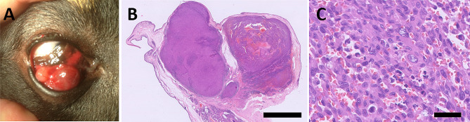
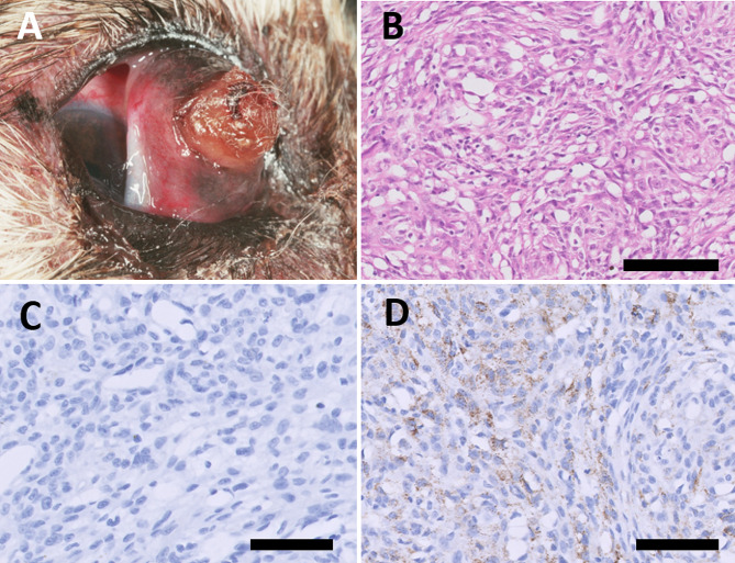
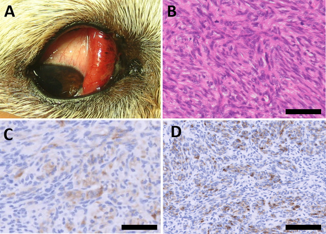
Results: The dogs presented were an 8-year-old Dachshund, a 12-year-old Akita and a 14-year-old Shetland Sheepdog. All three dogs were entire females. All three nictitating membrane tumours developed on the right nictitating membrane as firm or multilobulated hyperaemic masses. Two of the tumours were macroscopically nonpigmented, the third being partly pigmented on the surface and ulcerated. According to the histopathology and for two of the cases immunohistochemistry with dog-adapted protocols the diagnoses included one hemangiosarcoma and two amelanotic melanomas. Tumour regrowth developed in all three cases and repeated resections were completed 1, 2 and 3 times, respectively, with recurrence experienced within 1.5 months - 3 years.
Conclusions: Nonpigmented malignant spindle cell tumours of the canine membrana nictitans are rare. Treatment of choice should be complete excision with a minimal histologic tumour-free distance and in case of a recurrence a full resection of the nictitating membrane. We strongly recommend a dog-adapted protocol for immunohistochemistry.
期刊介绍:
Acta Veterinaria Scandinavica is an open access journal encompassing all aspects of veterinary research and medicine of domestic and wild animals.


 分享
分享
 求助内容:
求助内容: 应助结果提醒方式:
 扫码关注我们
