L. V. Kordyukova, A. V. Moiseenko, T. A. Timofeeva, I. T. Fedyakina
{"title":"使用升级版透射电子显微镜对包膜病毒进行冷冻电镜观察:甲型和乙型流感病毒以及 SARS-CoV-2","authors":"L. V. Kordyukova, A. V. Moiseenko, T. A. Timofeeva, I. T. Fedyakina","doi":"10.3103/s0096392523700153","DOIUrl":null,"url":null,"abstract":"<h3 data-test=\"abstract-sub-heading\">Abstract</h3><p>Cryo-electron microscopy (cryo-EM) is indispensable for the structural studies of enveloped viruses: dangerous pathogens of humans and animals. Yet, it requires highly specialized equipment as well as careful sample preparation. In this work, the capabilities of a JEOL JEM-2100 transmission electron microscope equipped with a cryo-transfer holder are used, and preliminary cryo-EM data for influenza A and B virus strains and SARS-CoV-2 inactivated with beta-propiolactone are presented. Image analysis allows us to (1) distinguish “empty” viral particles from “full” ones (containing nucleocapsid); (2) visualize the lipid bilayer of the viral envelope; (3) identify influenza virus surface antigens and the M1 protein layer combined with the inner lipid monolayer; and (4) distinguish different morphology of S-spikes on the surface of inactivated SARS-CoV-2 virions. The developed approach provides good image quality for both fundamental and applied research.</p>","PeriodicalId":19004,"journal":{"name":"Moscow University Biological Sciences Bulletin","volume":"16 1","pages":""},"PeriodicalIF":0.0000,"publicationDate":"2024-03-11","publicationTypes":"Journal Article","fieldsOfStudy":null,"isOpenAccess":false,"openAccessPdf":"","citationCount":"0","resultStr":"{\"title\":\"Cryo-Electron Microscopy of Enveloped Viruses Using an Upgraded Transmission Electron Microscope: Influenza Type A and B Viruses and SARS-CoV-2\",\"authors\":\"L. V. Kordyukova, A. V. Moiseenko, T. A. Timofeeva, I. T. Fedyakina\",\"doi\":\"10.3103/s0096392523700153\",\"DOIUrl\":null,\"url\":null,\"abstract\":\"<h3 data-test=\\\"abstract-sub-heading\\\">Abstract</h3><p>Cryo-electron microscopy (cryo-EM) is indispensable for the structural studies of enveloped viruses: dangerous pathogens of humans and animals. Yet, it requires highly specialized equipment as well as careful sample preparation. In this work, the capabilities of a JEOL JEM-2100 transmission electron microscope equipped with a cryo-transfer holder are used, and preliminary cryo-EM data for influenza A and B virus strains and SARS-CoV-2 inactivated with beta-propiolactone are presented. Image analysis allows us to (1) distinguish “empty” viral particles from “full” ones (containing nucleocapsid); (2) visualize the lipid bilayer of the viral envelope; (3) identify influenza virus surface antigens and the M1 protein layer combined with the inner lipid monolayer; and (4) distinguish different morphology of S-spikes on the surface of inactivated SARS-CoV-2 virions. The developed approach provides good image quality for both fundamental and applied research.</p>\",\"PeriodicalId\":19004,\"journal\":{\"name\":\"Moscow University Biological Sciences Bulletin\",\"volume\":\"16 1\",\"pages\":\"\"},\"PeriodicalIF\":0.0000,\"publicationDate\":\"2024-03-11\",\"publicationTypes\":\"Journal Article\",\"fieldsOfStudy\":null,\"isOpenAccess\":false,\"openAccessPdf\":\"\",\"citationCount\":\"0\",\"resultStr\":null,\"platform\":\"Semanticscholar\",\"paperid\":null,\"PeriodicalName\":\"Moscow University Biological Sciences Bulletin\",\"FirstCategoryId\":\"1085\",\"ListUrlMain\":\"https://doi.org/10.3103/s0096392523700153\",\"RegionNum\":0,\"RegionCategory\":null,\"ArticlePicture\":[],\"TitleCN\":null,\"AbstractTextCN\":null,\"PMCID\":null,\"EPubDate\":\"\",\"PubModel\":\"\",\"JCR\":\"Q3\",\"JCRName\":\"Agricultural and Biological Sciences\",\"Score\":null,\"Total\":0}","platform":"Semanticscholar","paperid":null,"PeriodicalName":"Moscow University Biological Sciences Bulletin","FirstCategoryId":"1085","ListUrlMain":"https://doi.org/10.3103/s0096392523700153","RegionNum":0,"RegionCategory":null,"ArticlePicture":[],"TitleCN":null,"AbstractTextCN":null,"PMCID":null,"EPubDate":"","PubModel":"","JCR":"Q3","JCRName":"Agricultural and Biological Sciences","Score":null,"Total":0}
Cryo-Electron Microscopy of Enveloped Viruses Using an Upgraded Transmission Electron Microscope: Influenza Type A and B Viruses and SARS-CoV-2
Abstract
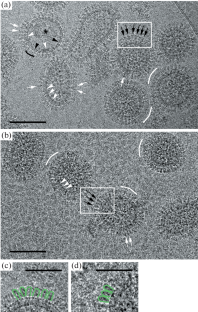
Cryo-electron microscopy (cryo-EM) is indispensable for the structural studies of enveloped viruses: dangerous pathogens of humans and animals. Yet, it requires highly specialized equipment as well as careful sample preparation. In this work, the capabilities of a JEOL JEM-2100 transmission electron microscope equipped with a cryo-transfer holder are used, and preliminary cryo-EM data for influenza A and B virus strains and SARS-CoV-2 inactivated with beta-propiolactone are presented. Image analysis allows us to (1) distinguish “empty” viral particles from “full” ones (containing nucleocapsid); (2) visualize the lipid bilayer of the viral envelope; (3) identify influenza virus surface antigens and the M1 protein layer combined with the inner lipid monolayer; and (4) distinguish different morphology of S-spikes on the surface of inactivated SARS-CoV-2 virions. The developed approach provides good image quality for both fundamental and applied research.
期刊介绍:
Moscow University Biological Sciences Bulletin is forum for research in all important areas of modern biology. It publishes original work on qualitative, analytical and experimental aspects of research. The scope of articles to be considered includes plant biology, zoology, ecology, evolutionary biology, biophysics, genetics, genomics, proteomics, molecular biology, cell biology, biochemistry, endocrinology, immunology, physiology, pharmacology, neuroscience, gerontology, developmental biology, bioinformatics, bioengineering, virology, and microbiology.


 分享
分享
 求助内容:
求助内容: 应助结果提醒方式:
 扫码关注我们
