{"title":"相关光镜和电子显微镜中的低温输送规程:从多层次成像到生物物理效应建模和 \"低温对抗 \"技术","authors":"O. V. Gradov","doi":"10.3103/s0096392523700244","DOIUrl":null,"url":null,"abstract":"<h3 data-test=\"abstract-sub-heading\">Abstract</h3><p>This paper is a technical and methodological note aimed to introduce into the practice of biological research methods a cryomicroscopy in a conveyor mode from small magnifications to the limits of magnification/resolution of scanning electron cryomicroscopy (CryoSEM). The protocol described can be applied to the samples of a low sample preparation complexity without ultratomy or processing typical of transmission electron microscopy. According to this protocol, samples are analyzed in a single microcuvette (chip) indexed by laboratory information management system and sequentially transferred from the nondestructive low-resolution optical microscopy instruments, such as lensless cryo-microscopes, to the CryoSEM/CryoESEM level (in programmable environments and atmospheres). Methods that were introduced and tested include correlative lensless cryomicroscopy and CryoSEM (including those with the sequential transition to microanalysis on wavelength-dispersive X-ray spectrometer on the Rowland circle) as well as microscopy and microinterferometry methods in the ranges from infrared to far-ultraviolet. Among the advantages of the cryoconveyor analysis protocols are sample preservation in a single portable cuvette-chip along with a possibility to establish spatial colocalization between data of optical and electron microscopy using pattern recognition software (all the way to indexing in the laboratory information system) in conducting a full range of microscopic examination. An opportunity is additionally provided for a comprehensive nondestructive sample analysis in a sequential study of microscopic systems with a possibility of variation at the subsequent stages of high-resolution microscopy, depending on the results obtained at the preceding stages of microscopy (at lower resolution).</p>","PeriodicalId":19004,"journal":{"name":"Moscow University Biological Sciences Bulletin","volume":"28 1","pages":""},"PeriodicalIF":0.0000,"publicationDate":"2024-03-11","publicationTypes":"Journal Article","fieldsOfStudy":null,"isOpenAccess":false,"openAccessPdf":"","citationCount":"0","resultStr":"{\"title\":\"Cryoconveyor Protocols in Correlative Light and Electron Microscopy: From Multilevel Imaging to Modeling the Biophysical Effects and “Cryotheranostics”\",\"authors\":\"O. V. Gradov\",\"doi\":\"10.3103/s0096392523700244\",\"DOIUrl\":null,\"url\":null,\"abstract\":\"<h3 data-test=\\\"abstract-sub-heading\\\">Abstract</h3><p>This paper is a technical and methodological note aimed to introduce into the practice of biological research methods a cryomicroscopy in a conveyor mode from small magnifications to the limits of magnification/resolution of scanning electron cryomicroscopy (CryoSEM). The protocol described can be applied to the samples of a low sample preparation complexity without ultratomy or processing typical of transmission electron microscopy. According to this protocol, samples are analyzed in a single microcuvette (chip) indexed by laboratory information management system and sequentially transferred from the nondestructive low-resolution optical microscopy instruments, such as lensless cryo-microscopes, to the CryoSEM/CryoESEM level (in programmable environments and atmospheres). Methods that were introduced and tested include correlative lensless cryomicroscopy and CryoSEM (including those with the sequential transition to microanalysis on wavelength-dispersive X-ray spectrometer on the Rowland circle) as well as microscopy and microinterferometry methods in the ranges from infrared to far-ultraviolet. Among the advantages of the cryoconveyor analysis protocols are sample preservation in a single portable cuvette-chip along with a possibility to establish spatial colocalization between data of optical and electron microscopy using pattern recognition software (all the way to indexing in the laboratory information system) in conducting a full range of microscopic examination. An opportunity is additionally provided for a comprehensive nondestructive sample analysis in a sequential study of microscopic systems with a possibility of variation at the subsequent stages of high-resolution microscopy, depending on the results obtained at the preceding stages of microscopy (at lower resolution).</p>\",\"PeriodicalId\":19004,\"journal\":{\"name\":\"Moscow University Biological Sciences Bulletin\",\"volume\":\"28 1\",\"pages\":\"\"},\"PeriodicalIF\":0.0000,\"publicationDate\":\"2024-03-11\",\"publicationTypes\":\"Journal Article\",\"fieldsOfStudy\":null,\"isOpenAccess\":false,\"openAccessPdf\":\"\",\"citationCount\":\"0\",\"resultStr\":null,\"platform\":\"Semanticscholar\",\"paperid\":null,\"PeriodicalName\":\"Moscow University Biological Sciences Bulletin\",\"FirstCategoryId\":\"1085\",\"ListUrlMain\":\"https://doi.org/10.3103/s0096392523700244\",\"RegionNum\":0,\"RegionCategory\":null,\"ArticlePicture\":[],\"TitleCN\":null,\"AbstractTextCN\":null,\"PMCID\":null,\"EPubDate\":\"\",\"PubModel\":\"\",\"JCR\":\"Q3\",\"JCRName\":\"Agricultural and Biological Sciences\",\"Score\":null,\"Total\":0}","platform":"Semanticscholar","paperid":null,"PeriodicalName":"Moscow University Biological Sciences Bulletin","FirstCategoryId":"1085","ListUrlMain":"https://doi.org/10.3103/s0096392523700244","RegionNum":0,"RegionCategory":null,"ArticlePicture":[],"TitleCN":null,"AbstractTextCN":null,"PMCID":null,"EPubDate":"","PubModel":"","JCR":"Q3","JCRName":"Agricultural and Biological Sciences","Score":null,"Total":0}
引用次数: 0
摘要
摘要 本文是一份技术和方法论说明,旨在将冷冻显微镜(CryoSEM)从小倍率到扫描电子冷冻显微镜(CryoSEM)放大倍率/分辨率极限的传输模式引入生物研究方法的实践中。所述方案适用于样品制备复杂度较低的样品,无需进行超微切片或典型的透射电子显微镜处理。根据该方案,样品在实验室信息管理系统索引的单个微量样品池(芯片)中进行分析,并按顺序从无损低分辨率光学显微镜仪器(如无镜头冷冻显微镜)转移到 CryoSEM/CryoESEM 级别(在可编程环境和气氛中)。引入和测试的方法包括相关的无透镜冷冻显微镜和 CryoSEM(包括在罗兰圈波长色散 X 射线光谱仪上依次过渡到微分析的方法),以及从红外线到远紫外线范围内的显微镜和微干涉测量方法。低温传送器分析协议的优点包括:样品保存在单个便携式比色皿芯片中,同时可以使用模式识别软件(一直到实验室信息系统中的索引)在光学和电子显微镜数据之间建立空间定位,以进行全方位的显微镜检查。此外,还提供了在显微系统的连续研究中进行全面无损样品分析的机会,根据前一阶段显微镜检查(分辨率较低)获得的结果,高分辨率显微镜检查的后续阶段有可能发生变化。
Cryoconveyor Protocols in Correlative Light and Electron Microscopy: From Multilevel Imaging to Modeling the Biophysical Effects and “Cryotheranostics”
Abstract
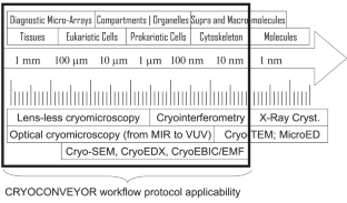
This paper is a technical and methodological note aimed to introduce into the practice of biological research methods a cryomicroscopy in a conveyor mode from small magnifications to the limits of magnification/resolution of scanning electron cryomicroscopy (CryoSEM). The protocol described can be applied to the samples of a low sample preparation complexity without ultratomy or processing typical of transmission electron microscopy. According to this protocol, samples are analyzed in a single microcuvette (chip) indexed by laboratory information management system and sequentially transferred from the nondestructive low-resolution optical microscopy instruments, such as lensless cryo-microscopes, to the CryoSEM/CryoESEM level (in programmable environments and atmospheres). Methods that were introduced and tested include correlative lensless cryomicroscopy and CryoSEM (including those with the sequential transition to microanalysis on wavelength-dispersive X-ray spectrometer on the Rowland circle) as well as microscopy and microinterferometry methods in the ranges from infrared to far-ultraviolet. Among the advantages of the cryoconveyor analysis protocols are sample preservation in a single portable cuvette-chip along with a possibility to establish spatial colocalization between data of optical and electron microscopy using pattern recognition software (all the way to indexing in the laboratory information system) in conducting a full range of microscopic examination. An opportunity is additionally provided for a comprehensive nondestructive sample analysis in a sequential study of microscopic systems with a possibility of variation at the subsequent stages of high-resolution microscopy, depending on the results obtained at the preceding stages of microscopy (at lower resolution).
期刊介绍:
Moscow University Biological Sciences Bulletin is forum for research in all important areas of modern biology. It publishes original work on qualitative, analytical and experimental aspects of research. The scope of articles to be considered includes plant biology, zoology, ecology, evolutionary biology, biophysics, genetics, genomics, proteomics, molecular biology, cell biology, biochemistry, endocrinology, immunology, physiology, pharmacology, neuroscience, gerontology, developmental biology, bioinformatics, bioengineering, virology, and microbiology.


 分享
分享
 求助内容:
求助内容: 应助结果提醒方式:
 扫码关注我们
