Christian Lehmann, Bertram Schmidt, Dietmar Stephan, Vera Meyer
{"title":"利用低真空扫描电子显微镜研究真菌菌丝复合建筑材料的界面。","authors":"Christian Lehmann, Bertram Schmidt, Dietmar Stephan, Vera Meyer","doi":"10.1111/jmi.13292","DOIUrl":null,"url":null,"abstract":"<p>Low-vacuum scanning electron microscopy (low-vacuum SEM) is widely used for different applications, such as the investigation of noncoated specimen or the observation of biological materials, which are not stable to high vacuum. In this study, the combination of mineral building materials (concrete or clay plaster) with a biological composite (fungal mycelium composite) by using low-vacuum SEM was investigated.</p><p>Fungal biotechnology is increasingly gaining prominence in addressing the challenges of sustainability transformation. The construction industry is one of the biggest contributors to the climate crises and, therefore, can highly profit from applications based on regenerative fungal materials. In this work, a fungal mycelium composite is used as alternative to conventional insulating materials like Styrofoam. However, to adapt bio-based products to the construction industry, investigations, optimisations and adaptations to existing solutions are needed. This paper examines the compatibility between fungal mycelium materials with mineral-based materials to demonstrate basic feasibility. For this purpose, fresh and hardened concrete specimens as well as clay plaster samples are combined with growing mycelium from the tinder fungus <i>Fomes fomentarius</i>. The contact zone between the mycelium composite and the mineral building materials is examined by scanning electron microscopy (SEM).</p><p>The combination of these materials proves to be feasible in general. The use of hardened concrete or clay with living mycelium composite appears to be the favoured variant, as the hyphae can grow into the surface of the building material and thus a layered structure with a stable connection is formed.</p><p>In order to work with the combination of low-density organic materials and higher-density inorganic materials simultaneously, low-vacuum SEM offers a suitable method to deliver results with reduced effort in preparation while maintaining high capture and magnification quality. Not only are image recordings possible with SE and BSE, but EDX measurements can also be carried out quickly without the influence of a coating. Depending on the signal used, as well as the magnification, image-recording strategies must be adapted. Especially when using SE, an image-integration method was used to reduce the build-up of point charges from the electron beam, which damages the mycelial hyphae. Additionally using different signals during image capture is recommended to confirm acquired information, avoiding misinterpretations.</p>","PeriodicalId":16484,"journal":{"name":"Journal of microscopy","volume":"294 2","pages":"203-214"},"PeriodicalIF":1.9000,"publicationDate":"2024-03-21","publicationTypes":"Journal Article","fieldsOfStudy":null,"isOpenAccess":false,"openAccessPdf":"https://onlinelibrary.wiley.com/doi/epdf/10.1111/jmi.13292","citationCount":"0","resultStr":"{\"title\":\"Investigation of the interface of fungal mycelium composite building materials by means of low-vacuum scanning electron microscopy\",\"authors\":\"Christian Lehmann, Bertram Schmidt, Dietmar Stephan, Vera Meyer\",\"doi\":\"10.1111/jmi.13292\",\"DOIUrl\":null,\"url\":null,\"abstract\":\"<p>Low-vacuum scanning electron microscopy (low-vacuum SEM) is widely used for different applications, such as the investigation of noncoated specimen or the observation of biological materials, which are not stable to high vacuum. In this study, the combination of mineral building materials (concrete or clay plaster) with a biological composite (fungal mycelium composite) by using low-vacuum SEM was investigated.</p><p>Fungal biotechnology is increasingly gaining prominence in addressing the challenges of sustainability transformation. The construction industry is one of the biggest contributors to the climate crises and, therefore, can highly profit from applications based on regenerative fungal materials. In this work, a fungal mycelium composite is used as alternative to conventional insulating materials like Styrofoam. However, to adapt bio-based products to the construction industry, investigations, optimisations and adaptations to existing solutions are needed. This paper examines the compatibility between fungal mycelium materials with mineral-based materials to demonstrate basic feasibility. For this purpose, fresh and hardened concrete specimens as well as clay plaster samples are combined with growing mycelium from the tinder fungus <i>Fomes fomentarius</i>. The contact zone between the mycelium composite and the mineral building materials is examined by scanning electron microscopy (SEM).</p><p>The combination of these materials proves to be feasible in general. The use of hardened concrete or clay with living mycelium composite appears to be the favoured variant, as the hyphae can grow into the surface of the building material and thus a layered structure with a stable connection is formed.</p><p>In order to work with the combination of low-density organic materials and higher-density inorganic materials simultaneously, low-vacuum SEM offers a suitable method to deliver results with reduced effort in preparation while maintaining high capture and magnification quality. Not only are image recordings possible with SE and BSE, but EDX measurements can also be carried out quickly without the influence of a coating. Depending on the signal used, as well as the magnification, image-recording strategies must be adapted. Especially when using SE, an image-integration method was used to reduce the build-up of point charges from the electron beam, which damages the mycelial hyphae. Additionally using different signals during image capture is recommended to confirm acquired information, avoiding misinterpretations.</p>\",\"PeriodicalId\":16484,\"journal\":{\"name\":\"Journal of microscopy\",\"volume\":\"294 2\",\"pages\":\"203-214\"},\"PeriodicalIF\":1.9000,\"publicationDate\":\"2024-03-21\",\"publicationTypes\":\"Journal Article\",\"fieldsOfStudy\":null,\"isOpenAccess\":false,\"openAccessPdf\":\"https://onlinelibrary.wiley.com/doi/epdf/10.1111/jmi.13292\",\"citationCount\":\"0\",\"resultStr\":null,\"platform\":\"Semanticscholar\",\"paperid\":null,\"PeriodicalName\":\"Journal of microscopy\",\"FirstCategoryId\":\"5\",\"ListUrlMain\":\"https://onlinelibrary.wiley.com/doi/10.1111/jmi.13292\",\"RegionNum\":4,\"RegionCategory\":\"工程技术\",\"ArticlePicture\":[],\"TitleCN\":null,\"AbstractTextCN\":null,\"PMCID\":null,\"EPubDate\":\"\",\"PubModel\":\"\",\"JCR\":\"Q3\",\"JCRName\":\"MICROSCOPY\",\"Score\":null,\"Total\":0}","platform":"Semanticscholar","paperid":null,"PeriodicalName":"Journal of microscopy","FirstCategoryId":"5","ListUrlMain":"https://onlinelibrary.wiley.com/doi/10.1111/jmi.13292","RegionNum":4,"RegionCategory":"工程技术","ArticlePicture":[],"TitleCN":null,"AbstractTextCN":null,"PMCID":null,"EPubDate":"","PubModel":"","JCR":"Q3","JCRName":"MICROSCOPY","Score":null,"Total":0}
引用次数: 0
摘要
低真空扫描电子显微镜(low-vacuum SEM)被广泛应用于不同领域,如研究非涂层试样或观察对高真空不稳定的生物材料。本研究利用低真空扫描电镜研究了矿物建筑材料(混凝土或粘土抹灰)与生物复合材料(真菌菌丝复合材料)的结合。真菌生物技术在应对可持续发展转型的挑战方面日益突出。建筑业是造成气候危机的最大因素之一,因此可以从基于再生真菌材料的应用中获益匪浅。在这项工作中,真菌菌丝复合材料被用来替代发泡胶等传统绝缘材料。然而,要将生物基产品应用于建筑行业,还需要对现有解决方案进行调查、优化和调整。本文研究了真菌菌丝体材料与矿物基材料之间的兼容性,以证明其基本可行性。为此,将新鲜和硬化的混凝土试样以及粘土灰泥样品与煤渣真菌 Fomes fomentarius 生长的菌丝结合在一起。通过扫描电子显微镜(SEM)检查了菌丝复合体与矿物建筑材料之间的接触区域。事实证明,这些材料的组合总体上是可行的。硬化混凝土或粘土与活菌丝复合体的使用似乎是最受欢迎的变体,因为菌丝可以长到建筑材料的表面,从而形成具有稳定连接的分层结构。为了同时处理低密度有机材料和高密度无机材料的组合,低真空扫描电子显微镜提供了一种合适的方法,在保持高捕捉和放大质量的同时,减少了准备工作的工作量。不仅可以使用 SE 和 BSE 进行图像记录,还可以在不受涂层影响的情况下快速进行 EDX 测量。根据所使用的信号和放大率,必须调整图像记录策略。特别是在使用 SE 时,需要使用图像整合方法来减少电子束产生的点电荷,因为点电荷会损坏菌丝菌丝。此外,建议在图像捕捉过程中使用不同的信号来确认获取的信息,避免误读。
Investigation of the interface of fungal mycelium composite building materials by means of low-vacuum scanning electron microscopy
Low-vacuum scanning electron microscopy (low-vacuum SEM) is widely used for different applications, such as the investigation of noncoated specimen or the observation of biological materials, which are not stable to high vacuum. In this study, the combination of mineral building materials (concrete or clay plaster) with a biological composite (fungal mycelium composite) by using low-vacuum SEM was investigated.
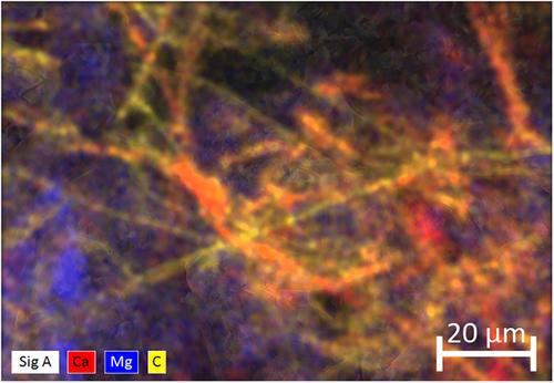
Fungal biotechnology is increasingly gaining prominence in addressing the challenges of sustainability transformation. The construction industry is one of the biggest contributors to the climate crises and, therefore, can highly profit from applications based on regenerative fungal materials. In this work, a fungal mycelium composite is used as alternative to conventional insulating materials like Styrofoam. However, to adapt bio-based products to the construction industry, investigations, optimisations and adaptations to existing solutions are needed. This paper examines the compatibility between fungal mycelium materials with mineral-based materials to demonstrate basic feasibility. For this purpose, fresh and hardened concrete specimens as well as clay plaster samples are combined with growing mycelium from the tinder fungus Fomes fomentarius. The contact zone between the mycelium composite and the mineral building materials is examined by scanning electron microscopy (SEM).
The combination of these materials proves to be feasible in general. The use of hardened concrete or clay with living mycelium composite appears to be the favoured variant, as the hyphae can grow into the surface of the building material and thus a layered structure with a stable connection is formed.
In order to work with the combination of low-density organic materials and higher-density inorganic materials simultaneously, low-vacuum SEM offers a suitable method to deliver results with reduced effort in preparation while maintaining high capture and magnification quality. Not only are image recordings possible with SE and BSE, but EDX measurements can also be carried out quickly without the influence of a coating. Depending on the signal used, as well as the magnification, image-recording strategies must be adapted. Especially when using SE, an image-integration method was used to reduce the build-up of point charges from the electron beam, which damages the mycelial hyphae. Additionally using different signals during image capture is recommended to confirm acquired information, avoiding misinterpretations.
期刊介绍:
The Journal of Microscopy is the oldest journal dedicated to the science of microscopy and the only peer-reviewed publication of the Royal Microscopical Society. It publishes papers that report on the very latest developments in microscopy such as advances in microscopy techniques or novel areas of application. The Journal does not seek to publish routine applications of microscopy or specimen preparation even though the submission may otherwise have a high scientific merit.
The scope covers research in the physical and biological sciences and covers imaging methods using light, electrons, X-rays and other radiations as well as atomic force and near field techniques. Interdisciplinary research is welcome. Papers pertaining to microscopy are also welcomed on optical theory, spectroscopy, novel specimen preparation and manipulation methods and image recording, processing and analysis including dynamic analysis of living specimens.
Publication types include full papers, hot topic fast tracked communications and review articles. Authors considering submitting a review article should contact the editorial office first.


 分享
分享
 求助内容:
求助内容: 应助结果提醒方式:
 扫码关注我们
