{"title":"使用抗坏血酸处理的肋软骨细胞衍生的无支架组织工程构建物促进关节软骨再生","authors":"Kaiwen Zheng, Yiyang Ma, Cheng Chiu, Mengxin Xue, Changqing Zhang, Dajiang Du","doi":"10.1016/j.jot.2024.02.005","DOIUrl":null,"url":null,"abstract":"<div><h3>Background</h3><p>Cartilage tissue engineering faces challenges related to the use of scaffolds and limited seed cells. This study aims to propose a cost-effective and straightforward approach using costal chondrocytes (CCs) as an alternative cell source to overcome these challenges, eliminating the need for special culture equipment or scaffolds.</p></div><div><h3>Methods</h3><p>CCs were cultured at a high cell density with and without ascorbic acid treatment, serving as the experimental and control groups, respectively. Viability and tissue-engineered constructs (TEC) formation were evaluated until day 14. Slices of TEC samples were used for histological staining to evaluate the secretion of glycosaminoglycans and different types of collagen proteins within the extracellular matrix. mRNA sequencing and qPCR were performed to examine gene expression related to cartilage matrix secretion in the chondrocytes. In vivo experiments were conducted by implanting TECs from different groups into the defect site, followed by sample collection after 12 weeks for histological staining and scoring to evaluate the extent of cartilage regeneration. Hematoxylin-eosin (HE), Safranin-O-Fast Green, and Masson's trichrome stainings were used to examine the content of cartilage-related matrix components in the in vivo repair tissue. Immunohistochemical staining for type I and type II collagen, as well as aggrecan, was performed to assess the presence and distribution of these specific markers. Additionally, immunohistochemical staining for type X collagen was used to observe any hypertrophic changes in the repaired tissue.</p></div><div><h3>Results</h3><p>Viability of the chondrocytes remained high throughout the culture period, and the TECs displayed an enriched extracellular matrix suitable for surgical procedures. In vitro study revealed glycosaminoglycan and type II collagen production in both groups of TEC, while the TEC matrix treated with ascorbic acid displayed greater abundance. The results of mRNA sequencing and qPCR showed that genes related to cartilage matrix secretion such as Sox9, Col2, and Acan were upregulated by ascorbic acid in costal chondrocytes. Although the addition of Asc-2P led to an increase in COL10 expression according to qPCR and RNA-seq results, the immunofluorescence staining results of the two groups of TECs exhibited similar distribution and fluorescence intensity. In vivo experiments showed that both groups of TEC could adhere to the defect sites and kept hyaline cartilage morphology until 12 weeks. TEC treated with ascorbic acid showed superior cartilage regeneration as evidenced by significantly higher ICRS and O'Driscoll scores and stronger Safranin-O and collagen staining mimicking native cartilage when compared to other groups. In addition, the immunohistochemical staining results of Collgan X indicated that, after 12 weeks, the ascorbic acid-treated TEC did not exhibit further hypertrophy upon transplantation into the defect site, but maintained an expression profile similar to untreated TECs, while slightly higher than the sham-operated group.</p></div><div><h3>Conclusion</h3><p>These results suggest that CC-derived scaffold-free TEC presents a promising method for articular cartilage regeneration. Ascorbic acid treatment enhances outcomes by promoting cartilage matrix production. This study provides valuable insights and potential advancements in the field of cartilage tissue engineering.</p></div><div><h3>The translational potential of this article</h3><p>Cartilage tissue engineering is an area of research with immense clinical potential. The approach presented in this article offers a cost-effective and straightforward solution, which can minimize the complexity of cell culture and scaffold fabrication. This simplification could offer several translational advantages, such as ease of use, rapid scalability, lower costs, and the potential for patient-specific clinical translation. The use of costal chondrocytes, which are easily obtainable, and the scaffold-free approach, which does not require specialized equipment or membranes, could be particularly advantageous in clinical settings, allowing for in situ regeneration of cartilage.</p></div>","PeriodicalId":16636,"journal":{"name":"Journal of Orthopaedic Translation","volume":"45 ","pages":"Pages 140-154"},"PeriodicalIF":9.8000,"publicationDate":"2024-03-01","publicationTypes":"Journal Article","fieldsOfStudy":null,"isOpenAccess":false,"openAccessPdf":"https://www.sciencedirect.com/science/article/pii/S2214031X24000214/pdfft?md5=b5653bc7717622e4f9f85e1876984c0c&pid=1-s2.0-S2214031X24000214-main.pdf","citationCount":"0","resultStr":"{\"title\":\"Enhanced articular cartilage regeneration using costal chondrocyte-derived scaffold-free tissue engineered constructs with ascorbic acid treatment\",\"authors\":\"Kaiwen Zheng, Yiyang Ma, Cheng Chiu, Mengxin Xue, Changqing Zhang, Dajiang Du\",\"doi\":\"10.1016/j.jot.2024.02.005\",\"DOIUrl\":null,\"url\":null,\"abstract\":\"<div><h3>Background</h3><p>Cartilage tissue engineering faces challenges related to the use of scaffolds and limited seed cells. This study aims to propose a cost-effective and straightforward approach using costal chondrocytes (CCs) as an alternative cell source to overcome these challenges, eliminating the need for special culture equipment or scaffolds.</p></div><div><h3>Methods</h3><p>CCs were cultured at a high cell density with and without ascorbic acid treatment, serving as the experimental and control groups, respectively. Viability and tissue-engineered constructs (TEC) formation were evaluated until day 14. Slices of TEC samples were used for histological staining to evaluate the secretion of glycosaminoglycans and different types of collagen proteins within the extracellular matrix. mRNA sequencing and qPCR were performed to examine gene expression related to cartilage matrix secretion in the chondrocytes. In vivo experiments were conducted by implanting TECs from different groups into the defect site, followed by sample collection after 12 weeks for histological staining and scoring to evaluate the extent of cartilage regeneration. Hematoxylin-eosin (HE), Safranin-O-Fast Green, and Masson's trichrome stainings were used to examine the content of cartilage-related matrix components in the in vivo repair tissue. Immunohistochemical staining for type I and type II collagen, as well as aggrecan, was performed to assess the presence and distribution of these specific markers. Additionally, immunohistochemical staining for type X collagen was used to observe any hypertrophic changes in the repaired tissue.</p></div><div><h3>Results</h3><p>Viability of the chondrocytes remained high throughout the culture period, and the TECs displayed an enriched extracellular matrix suitable for surgical procedures. In vitro study revealed glycosaminoglycan and type II collagen production in both groups of TEC, while the TEC matrix treated with ascorbic acid displayed greater abundance. The results of mRNA sequencing and qPCR showed that genes related to cartilage matrix secretion such as Sox9, Col2, and Acan were upregulated by ascorbic acid in costal chondrocytes. Although the addition of Asc-2P led to an increase in COL10 expression according to qPCR and RNA-seq results, the immunofluorescence staining results of the two groups of TECs exhibited similar distribution and fluorescence intensity. In vivo experiments showed that both groups of TEC could adhere to the defect sites and kept hyaline cartilage morphology until 12 weeks. TEC treated with ascorbic acid showed superior cartilage regeneration as evidenced by significantly higher ICRS and O'Driscoll scores and stronger Safranin-O and collagen staining mimicking native cartilage when compared to other groups. In addition, the immunohistochemical staining results of Collgan X indicated that, after 12 weeks, the ascorbic acid-treated TEC did not exhibit further hypertrophy upon transplantation into the defect site, but maintained an expression profile similar to untreated TECs, while slightly higher than the sham-operated group.</p></div><div><h3>Conclusion</h3><p>These results suggest that CC-derived scaffold-free TEC presents a promising method for articular cartilage regeneration. Ascorbic acid treatment enhances outcomes by promoting cartilage matrix production. This study provides valuable insights and potential advancements in the field of cartilage tissue engineering.</p></div><div><h3>The translational potential of this article</h3><p>Cartilage tissue engineering is an area of research with immense clinical potential. The approach presented in this article offers a cost-effective and straightforward solution, which can minimize the complexity of cell culture and scaffold fabrication. This simplification could offer several translational advantages, such as ease of use, rapid scalability, lower costs, and the potential for patient-specific clinical translation. The use of costal chondrocytes, which are easily obtainable, and the scaffold-free approach, which does not require specialized equipment or membranes, could be particularly advantageous in clinical settings, allowing for in situ regeneration of cartilage.</p></div>\",\"PeriodicalId\":16636,\"journal\":{\"name\":\"Journal of Orthopaedic Translation\",\"volume\":\"45 \",\"pages\":\"Pages 140-154\"},\"PeriodicalIF\":9.8000,\"publicationDate\":\"2024-03-01\",\"publicationTypes\":\"Journal Article\",\"fieldsOfStudy\":null,\"isOpenAccess\":false,\"openAccessPdf\":\"https://www.sciencedirect.com/science/article/pii/S2214031X24000214/pdfft?md5=b5653bc7717622e4f9f85e1876984c0c&pid=1-s2.0-S2214031X24000214-main.pdf\",\"citationCount\":\"0\",\"resultStr\":null,\"platform\":\"Semanticscholar\",\"paperid\":null,\"PeriodicalName\":\"Journal of Orthopaedic Translation\",\"FirstCategoryId\":\"3\",\"ListUrlMain\":\"https://www.sciencedirect.com/science/article/pii/S2214031X24000214\",\"RegionNum\":1,\"RegionCategory\":\"医学\",\"ArticlePicture\":[],\"TitleCN\":null,\"AbstractTextCN\":null,\"PMCID\":null,\"EPubDate\":\"2024/3/22 0:00:00\",\"PubModel\":\"Epub\",\"JCR\":\"Q1\",\"JCRName\":\"ORTHOPEDICS\",\"Score\":null,\"Total\":0}","platform":"Semanticscholar","paperid":null,"PeriodicalName":"Journal of Orthopaedic Translation","FirstCategoryId":"3","ListUrlMain":"https://www.sciencedirect.com/science/article/pii/S2214031X24000214","RegionNum":1,"RegionCategory":"医学","ArticlePicture":[],"TitleCN":null,"AbstractTextCN":null,"PMCID":null,"EPubDate":"2024/3/22 0:00:00","PubModel":"Epub","JCR":"Q1","JCRName":"ORTHOPEDICS","Score":null,"Total":0}
Enhanced articular cartilage regeneration using costal chondrocyte-derived scaffold-free tissue engineered constructs with ascorbic acid treatment
Background
Cartilage tissue engineering faces challenges related to the use of scaffolds and limited seed cells. This study aims to propose a cost-effective and straightforward approach using costal chondrocytes (CCs) as an alternative cell source to overcome these challenges, eliminating the need for special culture equipment or scaffolds.
Methods
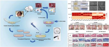
CCs were cultured at a high cell density with and without ascorbic acid treatment, serving as the experimental and control groups, respectively. Viability and tissue-engineered constructs (TEC) formation were evaluated until day 14. Slices of TEC samples were used for histological staining to evaluate the secretion of glycosaminoglycans and different types of collagen proteins within the extracellular matrix. mRNA sequencing and qPCR were performed to examine gene expression related to cartilage matrix secretion in the chondrocytes. In vivo experiments were conducted by implanting TECs from different groups into the defect site, followed by sample collection after 12 weeks for histological staining and scoring to evaluate the extent of cartilage regeneration. Hematoxylin-eosin (HE), Safranin-O-Fast Green, and Masson's trichrome stainings were used to examine the content of cartilage-related matrix components in the in vivo repair tissue. Immunohistochemical staining for type I and type II collagen, as well as aggrecan, was performed to assess the presence and distribution of these specific markers. Additionally, immunohistochemical staining for type X collagen was used to observe any hypertrophic changes in the repaired tissue.
Results
Viability of the chondrocytes remained high throughout the culture period, and the TECs displayed an enriched extracellular matrix suitable for surgical procedures. In vitro study revealed glycosaminoglycan and type II collagen production in both groups of TEC, while the TEC matrix treated with ascorbic acid displayed greater abundance. The results of mRNA sequencing and qPCR showed that genes related to cartilage matrix secretion such as Sox9, Col2, and Acan were upregulated by ascorbic acid in costal chondrocytes. Although the addition of Asc-2P led to an increase in COL10 expression according to qPCR and RNA-seq results, the immunofluorescence staining results of the two groups of TECs exhibited similar distribution and fluorescence intensity. In vivo experiments showed that both groups of TEC could adhere to the defect sites and kept hyaline cartilage morphology until 12 weeks. TEC treated with ascorbic acid showed superior cartilage regeneration as evidenced by significantly higher ICRS and O'Driscoll scores and stronger Safranin-O and collagen staining mimicking native cartilage when compared to other groups. In addition, the immunohistochemical staining results of Collgan X indicated that, after 12 weeks, the ascorbic acid-treated TEC did not exhibit further hypertrophy upon transplantation into the defect site, but maintained an expression profile similar to untreated TECs, while slightly higher than the sham-operated group.
Conclusion
These results suggest that CC-derived scaffold-free TEC presents a promising method for articular cartilage regeneration. Ascorbic acid treatment enhances outcomes by promoting cartilage matrix production. This study provides valuable insights and potential advancements in the field of cartilage tissue engineering.
The translational potential of this article
Cartilage tissue engineering is an area of research with immense clinical potential. The approach presented in this article offers a cost-effective and straightforward solution, which can minimize the complexity of cell culture and scaffold fabrication. This simplification could offer several translational advantages, such as ease of use, rapid scalability, lower costs, and the potential for patient-specific clinical translation. The use of costal chondrocytes, which are easily obtainable, and the scaffold-free approach, which does not require specialized equipment or membranes, could be particularly advantageous in clinical settings, allowing for in situ regeneration of cartilage.
期刊介绍:
The Journal of Orthopaedic Translation (JOT) is the official peer-reviewed, open access journal of the Chinese Speaking Orthopaedic Society (CSOS) and the International Chinese Musculoskeletal Research Society (ICMRS). It is published quarterly, in January, April, July and October, by Elsevier.


 分享
分享
 求助内容:
求助内容: 应助结果提醒方式:
 扫码关注我们
