Jun Ma, Ronald Xie, Shamini Ayyadhury, Cheng Ge, Anubha Gupta, Ritu Gupta, Song Gu, Yao Zhang, Gihun Lee, Joonkee Kim, Wei Lou, Haofeng Li, Eric Upschulte, Timo Dickscheid, José Guilherme de Almeida, Yixin Wang, Lin Han, Xin Yang, Marco Labagnara, Vojislav Gligorovski, Maxime Scheder, Sahand Jamal Rahi, Carly Kempster, Alice Pollitt, Leon Espinosa, Tâm Mignot, Jan Moritz Middeke, Jan-Niklas Eckardt, Wangkai Li, Zhaoyang Li, Xiaochen Cai, Bizhe Bai, Noah F. Greenwald, David Van Valen, Erin Weisbart, Beth A. Cimini, Trevor Cheung, Oscar Brück, Gary D. Bader, Bo Wang
{"title":"多模态细胞分割挑战:走向通用解决方案。","authors":"Jun Ma, Ronald Xie, Shamini Ayyadhury, Cheng Ge, Anubha Gupta, Ritu Gupta, Song Gu, Yao Zhang, Gihun Lee, Joonkee Kim, Wei Lou, Haofeng Li, Eric Upschulte, Timo Dickscheid, José Guilherme de Almeida, Yixin Wang, Lin Han, Xin Yang, Marco Labagnara, Vojislav Gligorovski, Maxime Scheder, Sahand Jamal Rahi, Carly Kempster, Alice Pollitt, Leon Espinosa, Tâm Mignot, Jan Moritz Middeke, Jan-Niklas Eckardt, Wangkai Li, Zhaoyang Li, Xiaochen Cai, Bizhe Bai, Noah F. Greenwald, David Van Valen, Erin Weisbart, Beth A. Cimini, Trevor Cheung, Oscar Brück, Gary D. Bader, Bo Wang","doi":"10.1038/s41592-024-02233-6","DOIUrl":null,"url":null,"abstract":"Cell segmentation is a critical step for quantitative single-cell analysis in microscopy images. Existing cell segmentation methods are often tailored to specific modalities or require manual interventions to specify hyper-parameters in different experimental settings. Here, we present a multimodality cell segmentation benchmark, comprising more than 1,500 labeled images derived from more than 50 diverse biological experiments. The top participants developed a Transformer-based deep-learning algorithm that not only exceeds existing methods but can also be applied to diverse microscopy images across imaging platforms and tissue types without manual parameter adjustments. This benchmark and the improved algorithm offer promising avenues for more accurate and versatile cell analysis in microscopy imaging. Cell segmentation is crucial in many image analysis pipelines. This analysis compares many tools on a multimodal cell segmentation benchmark. A Transformer-based model performed best in terms of performance and general applicability.","PeriodicalId":18981,"journal":{"name":"Nature Methods","volume":"21 6","pages":"1103-1113"},"PeriodicalIF":32.1000,"publicationDate":"2024-03-26","publicationTypes":"Journal Article","fieldsOfStudy":null,"isOpenAccess":false,"openAccessPdf":"","citationCount":"0","resultStr":"{\"title\":\"The multimodality cell segmentation challenge: toward universal solutions\",\"authors\":\"Jun Ma, Ronald Xie, Shamini Ayyadhury, Cheng Ge, Anubha Gupta, Ritu Gupta, Song Gu, Yao Zhang, Gihun Lee, Joonkee Kim, Wei Lou, Haofeng Li, Eric Upschulte, Timo Dickscheid, José Guilherme de Almeida, Yixin Wang, Lin Han, Xin Yang, Marco Labagnara, Vojislav Gligorovski, Maxime Scheder, Sahand Jamal Rahi, Carly Kempster, Alice Pollitt, Leon Espinosa, Tâm Mignot, Jan Moritz Middeke, Jan-Niklas Eckardt, Wangkai Li, Zhaoyang Li, Xiaochen Cai, Bizhe Bai, Noah F. Greenwald, David Van Valen, Erin Weisbart, Beth A. Cimini, Trevor Cheung, Oscar Brück, Gary D. Bader, Bo Wang\",\"doi\":\"10.1038/s41592-024-02233-6\",\"DOIUrl\":null,\"url\":null,\"abstract\":\"Cell segmentation is a critical step for quantitative single-cell analysis in microscopy images. Existing cell segmentation methods are often tailored to specific modalities or require manual interventions to specify hyper-parameters in different experimental settings. Here, we present a multimodality cell segmentation benchmark, comprising more than 1,500 labeled images derived from more than 50 diverse biological experiments. The top participants developed a Transformer-based deep-learning algorithm that not only exceeds existing methods but can also be applied to diverse microscopy images across imaging platforms and tissue types without manual parameter adjustments. This benchmark and the improved algorithm offer promising avenues for more accurate and versatile cell analysis in microscopy imaging. Cell segmentation is crucial in many image analysis pipelines. This analysis compares many tools on a multimodal cell segmentation benchmark. A Transformer-based model performed best in terms of performance and general applicability.\",\"PeriodicalId\":18981,\"journal\":{\"name\":\"Nature Methods\",\"volume\":\"21 6\",\"pages\":\"1103-1113\"},\"PeriodicalIF\":32.1000,\"publicationDate\":\"2024-03-26\",\"publicationTypes\":\"Journal Article\",\"fieldsOfStudy\":null,\"isOpenAccess\":false,\"openAccessPdf\":\"\",\"citationCount\":\"0\",\"resultStr\":null,\"platform\":\"Semanticscholar\",\"paperid\":null,\"PeriodicalName\":\"Nature Methods\",\"FirstCategoryId\":\"99\",\"ListUrlMain\":\"https://www.nature.com/articles/s41592-024-02233-6\",\"RegionNum\":1,\"RegionCategory\":\"生物学\",\"ArticlePicture\":[],\"TitleCN\":null,\"AbstractTextCN\":null,\"PMCID\":null,\"EPubDate\":\"\",\"PubModel\":\"\",\"JCR\":\"Q1\",\"JCRName\":\"BIOCHEMICAL RESEARCH METHODS\",\"Score\":null,\"Total\":0}","platform":"Semanticscholar","paperid":null,"PeriodicalName":"Nature Methods","FirstCategoryId":"99","ListUrlMain":"https://www.nature.com/articles/s41592-024-02233-6","RegionNum":1,"RegionCategory":"生物学","ArticlePicture":[],"TitleCN":null,"AbstractTextCN":null,"PMCID":null,"EPubDate":"","PubModel":"","JCR":"Q1","JCRName":"BIOCHEMICAL RESEARCH METHODS","Score":null,"Total":0}
The multimodality cell segmentation challenge: toward universal solutions
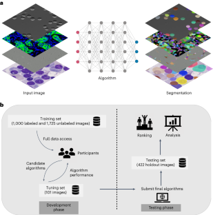
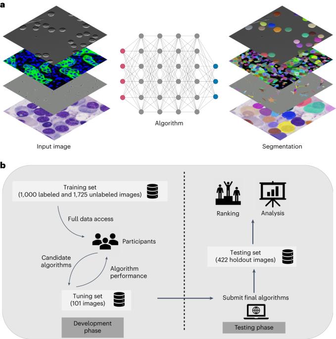
Cell segmentation is a critical step for quantitative single-cell analysis in microscopy images. Existing cell segmentation methods are often tailored to specific modalities or require manual interventions to specify hyper-parameters in different experimental settings. Here, we present a multimodality cell segmentation benchmark, comprising more than 1,500 labeled images derived from more than 50 diverse biological experiments. The top participants developed a Transformer-based deep-learning algorithm that not only exceeds existing methods but can also be applied to diverse microscopy images across imaging platforms and tissue types without manual parameter adjustments. This benchmark and the improved algorithm offer promising avenues for more accurate and versatile cell analysis in microscopy imaging. Cell segmentation is crucial in many image analysis pipelines. This analysis compares many tools on a multimodal cell segmentation benchmark. A Transformer-based model performed best in terms of performance and general applicability.
期刊介绍:
Nature Methods is a monthly journal that focuses on publishing innovative methods and substantial enhancements to fundamental life sciences research techniques. Geared towards a diverse, interdisciplinary readership of researchers in academia and industry engaged in laboratory work, the journal offers new tools for research and emphasizes the immediate practical significance of the featured work. It publishes primary research papers and reviews recent technical and methodological advancements, with a particular interest in primary methods papers relevant to the biological and biomedical sciences. This includes methods rooted in chemistry with practical applications for studying biological problems.


 分享
分享
 求助内容:
求助内容: 应助结果提醒方式:
 扫码关注我们
