Md Nasful Huda Prince, Benjamin Garcia, Cory Henn, Yating Yi, Etsuo A. Susaki, Yuki Watakabe, Tomomi Nemoto, Keith A. Lidke, Hu Zhao, Irene Salinas Remiro, Sheng Liu, Tonmoy Chakraborty
{"title":"用于大型组织成像的信号改进型超快光片显微镜","authors":"Md Nasful Huda Prince, Benjamin Garcia, Cory Henn, Yating Yi, Etsuo A. Susaki, Yuki Watakabe, Tomomi Nemoto, Keith A. Lidke, Hu Zhao, Irene Salinas Remiro, Sheng Liu, Tonmoy Chakraborty","doi":"10.1038/s44172-024-00205-4","DOIUrl":null,"url":null,"abstract":"Axially swept light-sheet microscope in conjunction with tissue clearing enables three-dimensional morphological investigation of millimeter-scaled tissues at isotropic sub-micron resolution. However, these microscopes suffer from low detection signal and slow imaging speed. Here we report a simple and efficient imaging platform that employs precise control of two fixed distant light-sheet foci for axial sweeping. This enables full field of view imaging at 40 frames per second, a four-fold improvement over the current state-of-the-art. In addition, in a particular frame rate, our method doubles the signal compared to the existing techniques. To augment the overall imaging performance, we also developed a deep learning based tissue information classifier that enables faster determination of tissue boundary. We demonstrated the performance of our imaging platform on various cleared tissue samples and delineated its robustness over a wide range of clearing protocols. Md Nasful Huda Prince and colleagues propose a tissue imaging system with isotropic sub-micron resolution. The method intelligently delineates tissue borders and captures images faster and with enhanced signal quality.","PeriodicalId":72644,"journal":{"name":"Communications engineering","volume":" ","pages":"1-13"},"PeriodicalIF":0.0000,"publicationDate":"2024-04-02","publicationTypes":"Journal Article","fieldsOfStudy":null,"isOpenAccess":false,"openAccessPdf":"https://www.nature.com/articles/s44172-024-00205-4.pdf","citationCount":"0","resultStr":"{\"title\":\"Signal improved ultra-fast light-sheet microscope for large tissue imaging\",\"authors\":\"Md Nasful Huda Prince, Benjamin Garcia, Cory Henn, Yating Yi, Etsuo A. Susaki, Yuki Watakabe, Tomomi Nemoto, Keith A. Lidke, Hu Zhao, Irene Salinas Remiro, Sheng Liu, Tonmoy Chakraborty\",\"doi\":\"10.1038/s44172-024-00205-4\",\"DOIUrl\":null,\"url\":null,\"abstract\":\"Axially swept light-sheet microscope in conjunction with tissue clearing enables three-dimensional morphological investigation of millimeter-scaled tissues at isotropic sub-micron resolution. However, these microscopes suffer from low detection signal and slow imaging speed. Here we report a simple and efficient imaging platform that employs precise control of two fixed distant light-sheet foci for axial sweeping. This enables full field of view imaging at 40 frames per second, a four-fold improvement over the current state-of-the-art. In addition, in a particular frame rate, our method doubles the signal compared to the existing techniques. To augment the overall imaging performance, we also developed a deep learning based tissue information classifier that enables faster determination of tissue boundary. We demonstrated the performance of our imaging platform on various cleared tissue samples and delineated its robustness over a wide range of clearing protocols. Md Nasful Huda Prince and colleagues propose a tissue imaging system with isotropic sub-micron resolution. The method intelligently delineates tissue borders and captures images faster and with enhanced signal quality.\",\"PeriodicalId\":72644,\"journal\":{\"name\":\"Communications engineering\",\"volume\":\" \",\"pages\":\"1-13\"},\"PeriodicalIF\":0.0000,\"publicationDate\":\"2024-04-02\",\"publicationTypes\":\"Journal Article\",\"fieldsOfStudy\":null,\"isOpenAccess\":false,\"openAccessPdf\":\"https://www.nature.com/articles/s44172-024-00205-4.pdf\",\"citationCount\":\"0\",\"resultStr\":null,\"platform\":\"Semanticscholar\",\"paperid\":null,\"PeriodicalName\":\"Communications engineering\",\"FirstCategoryId\":\"1085\",\"ListUrlMain\":\"https://www.nature.com/articles/s44172-024-00205-4\",\"RegionNum\":0,\"RegionCategory\":null,\"ArticlePicture\":[],\"TitleCN\":null,\"AbstractTextCN\":null,\"PMCID\":null,\"EPubDate\":\"\",\"PubModel\":\"\",\"JCR\":\"\",\"JCRName\":\"\",\"Score\":null,\"Total\":0}","platform":"Semanticscholar","paperid":null,"PeriodicalName":"Communications engineering","FirstCategoryId":"1085","ListUrlMain":"https://www.nature.com/articles/s44172-024-00205-4","RegionNum":0,"RegionCategory":null,"ArticlePicture":[],"TitleCN":null,"AbstractTextCN":null,"PMCID":null,"EPubDate":"","PubModel":"","JCR":"","JCRName":"","Score":null,"Total":0}
引用次数: 0
摘要
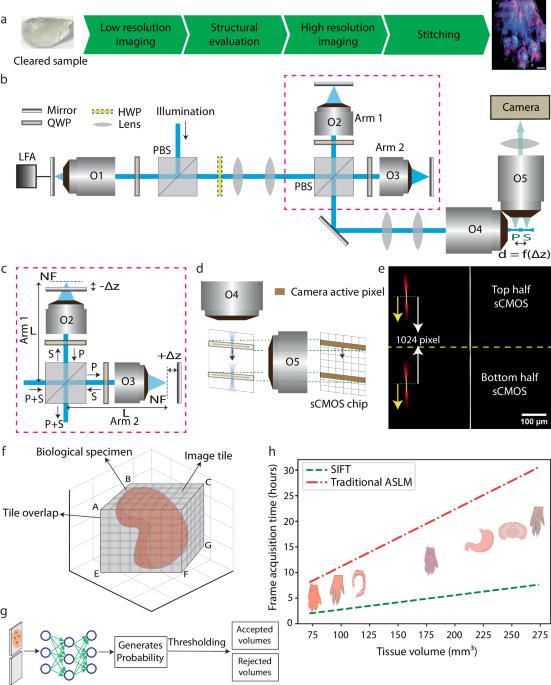
轴向扫描光片显微镜与组织清除技术相结合,能够以各向同性的亚微米分辨率对毫米级组织进行三维形态学研究。然而,这些显微镜存在检测信号低和成像速度慢的问题。在此,我们报告了一种简单而高效的成像平台,它采用精确控制两个固定的远距离光片焦点来进行轴向扫描。这样就能以每秒 40 帧的速度进行全视场成像,比目前最先进的技术提高了四倍。此外,与现有技术相比,我们的方法在特定帧频下可将信号增加一倍。为了提高整体成像性能,我们还开发了基于深度学习的组织信息分类器,能够更快地确定组织边界。我们在各种清除过的组织样本上演示了我们的成像平台的性能,并描述了它在各种清除协议中的稳健性。Md Nasful Huda Prince 及其同事提出了一种具有各向同性亚微米分辨率的组织成像系统。该方法能智能地划定组织边界,捕捉图像的速度更快,信号质量更高。
Signal improved ultra-fast light-sheet microscope for large tissue imaging
Axially swept light-sheet microscope in conjunction with tissue clearing enables three-dimensional morphological investigation of millimeter-scaled tissues at isotropic sub-micron resolution. However, these microscopes suffer from low detection signal and slow imaging speed. Here we report a simple and efficient imaging platform that employs precise control of two fixed distant light-sheet foci for axial sweeping. This enables full field of view imaging at 40 frames per second, a four-fold improvement over the current state-of-the-art. In addition, in a particular frame rate, our method doubles the signal compared to the existing techniques. To augment the overall imaging performance, we also developed a deep learning based tissue information classifier that enables faster determination of tissue boundary. We demonstrated the performance of our imaging platform on various cleared tissue samples and delineated its robustness over a wide range of clearing protocols. Md Nasful Huda Prince and colleagues propose a tissue imaging system with isotropic sub-micron resolution. The method intelligently delineates tissue borders and captures images faster and with enhanced signal quality.


 分享
分享

 求助内容:
求助内容: 应助结果提醒方式:
 扫码关注我们
