Elizabeth Lea Schmidt, Zihao Ou, Erving Ximendes, Han Cui, Carl H. C. Keck, Daniel Jaque, Guosong Hong
{"title":"近红外 II 荧光成像","authors":"Elizabeth Lea Schmidt, Zihao Ou, Erving Ximendes, Han Cui, Carl H. C. Keck, Daniel Jaque, Guosong Hong","doi":"10.1038/s43586-024-00301-x","DOIUrl":null,"url":null,"abstract":"Fluorescence imaging in the second near-infrared (NIR-II) window enables deep-tissue imaging with high resolution and improved contrast by taking advantage of the reduced light scattering and tissue autofluorescence in this region of the spectrum. NIR-II fluorescence imaging uses photoluminescent contrast agents — including carbon nanotubes, quantum dots, rare earth-doped nanocrystals, gold nanoclusters, small molecules and their aggregates — and fluorescent proteins, which all exhibit fluorescence in the 1,000–3,000 nm range. After administration of these fluorophores in vivo, live animals can be imaged with specialized detectors and optical instruments, yielding images with contrast and resolution unparalleled by conventional visible and near-infrared fluorescence imaging. This powerful approach enables dynamic imaging of vascular structures and haemodynamics; molecular imaging and image-guided surgery of tumours; and visualization of deep-seated structures, such as the gastrointestinal system. NIR-II fluorescence imaging has revolutionized biomedical imaging over the past 15 years and is poised to make comparable advancements in cardiology, neurobiology and gastroenterology. This Primer describes the principles of NIR-II fluorescence imaging, reviews the most used fluorophores, outlines implementation approaches and discusses specific scientific and clinical applications. Furthermore, the limitations of NIR-II fluorescence imaging are addressed and future opportunities across various scientific domains are explored. Deep tissues can be imaged with high resolution and greater contrast by performing fluorescence imaging in the second near-infrared (NIR-II) window. This Primer summarizes how NIR-II fluorescence imaging can be used in animal models, exploring commonly used fluorophores and implementation approaches across a range of scientific and clinical applications.","PeriodicalId":74250,"journal":{"name":"Nature reviews. Methods primers","volume":" ","pages":"1-22"},"PeriodicalIF":50.1000,"publicationDate":"2024-04-04","publicationTypes":"Journal Article","fieldsOfStudy":null,"isOpenAccess":false,"openAccessPdf":"","citationCount":"0","resultStr":"{\"title\":\"Near-infrared II fluorescence imaging\",\"authors\":\"Elizabeth Lea Schmidt, Zihao Ou, Erving Ximendes, Han Cui, Carl H. C. Keck, Daniel Jaque, Guosong Hong\",\"doi\":\"10.1038/s43586-024-00301-x\",\"DOIUrl\":null,\"url\":null,\"abstract\":\"Fluorescence imaging in the second near-infrared (NIR-II) window enables deep-tissue imaging with high resolution and improved contrast by taking advantage of the reduced light scattering and tissue autofluorescence in this region of the spectrum. NIR-II fluorescence imaging uses photoluminescent contrast agents — including carbon nanotubes, quantum dots, rare earth-doped nanocrystals, gold nanoclusters, small molecules and their aggregates — and fluorescent proteins, which all exhibit fluorescence in the 1,000–3,000 nm range. After administration of these fluorophores in vivo, live animals can be imaged with specialized detectors and optical instruments, yielding images with contrast and resolution unparalleled by conventional visible and near-infrared fluorescence imaging. This powerful approach enables dynamic imaging of vascular structures and haemodynamics; molecular imaging and image-guided surgery of tumours; and visualization of deep-seated structures, such as the gastrointestinal system. NIR-II fluorescence imaging has revolutionized biomedical imaging over the past 15 years and is poised to make comparable advancements in cardiology, neurobiology and gastroenterology. This Primer describes the principles of NIR-II fluorescence imaging, reviews the most used fluorophores, outlines implementation approaches and discusses specific scientific and clinical applications. Furthermore, the limitations of NIR-II fluorescence imaging are addressed and future opportunities across various scientific domains are explored. Deep tissues can be imaged with high resolution and greater contrast by performing fluorescence imaging in the second near-infrared (NIR-II) window. This Primer summarizes how NIR-II fluorescence imaging can be used in animal models, exploring commonly used fluorophores and implementation approaches across a range of scientific and clinical applications.\",\"PeriodicalId\":74250,\"journal\":{\"name\":\"Nature reviews. Methods primers\",\"volume\":\" \",\"pages\":\"1-22\"},\"PeriodicalIF\":50.1000,\"publicationDate\":\"2024-04-04\",\"publicationTypes\":\"Journal Article\",\"fieldsOfStudy\":null,\"isOpenAccess\":false,\"openAccessPdf\":\"\",\"citationCount\":\"0\",\"resultStr\":null,\"platform\":\"Semanticscholar\",\"paperid\":null,\"PeriodicalName\":\"Nature reviews. Methods primers\",\"FirstCategoryId\":\"1085\",\"ListUrlMain\":\"https://www.nature.com/articles/s43586-024-00301-x\",\"RegionNum\":0,\"RegionCategory\":null,\"ArticlePicture\":[],\"TitleCN\":null,\"AbstractTextCN\":null,\"PMCID\":null,\"EPubDate\":\"\",\"PubModel\":\"\",\"JCR\":\"Q1\",\"JCRName\":\"MULTIDISCIPLINARY SCIENCES\",\"Score\":null,\"Total\":0}","platform":"Semanticscholar","paperid":null,"PeriodicalName":"Nature reviews. Methods primers","FirstCategoryId":"1085","ListUrlMain":"https://www.nature.com/articles/s43586-024-00301-x","RegionNum":0,"RegionCategory":null,"ArticlePicture":[],"TitleCN":null,"AbstractTextCN":null,"PMCID":null,"EPubDate":"","PubModel":"","JCR":"Q1","JCRName":"MULTIDISCIPLINARY SCIENCES","Score":null,"Total":0}
引用次数: 0
摘要
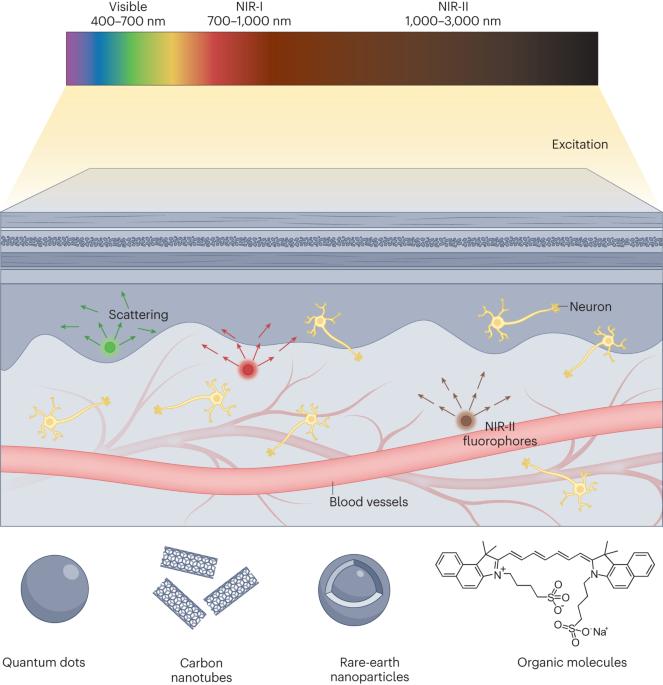
利用第二近红外(NIR-II)窗口的荧光成像技术,可以利用该光谱区域较低的光散射和组织自发荧光,进行高分辨率和高对比度的深部组织成像。近红外-II 荧光成像使用的光致发光造影剂包括碳纳米管、量子点、掺稀土纳米晶体、金纳米团簇、小分子及其聚集体和荧光蛋白,它们都在 1000-3000 纳米范围内发出荧光。在体内施用这些荧光团后,活体动物可以通过专门的探测器和光学仪器成像,从而获得对比度和分辨率都无与伦比的传统可见光和近红外荧光成像图像。这种强大的方法可实现血管结构和血液动力学的动态成像;肿瘤的分子成像和图像引导手术;以及胃肠系统等深层结构的可视化。近红外-II荧光成像技术在过去15年中彻底改变了生物医学成像技术,并有望在心脏病学、神经生物学和胃肠病学领域取得类似进展。本入门指南介绍了近红外 II 荧光成像的原理,回顾了最常用的荧光团,概述了实施方法,并讨论了具体的科学和临床应用。此外,还讨论了近红外-II 荧光成像的局限性,并探讨了各个科学领域的未来机遇。通过在第二个近红外(NIR-II)窗口进行荧光成像,可以对深部组织进行高分辨率和高对比度成像。本入门指南总结了如何在动物模型中使用近红外-II 荧光成像,探讨了一系列科学和临床应用中常用的荧光团和实施方法。
Fluorescence imaging in the second near-infrared (NIR-II) window enables deep-tissue imaging with high resolution and improved contrast by taking advantage of the reduced light scattering and tissue autofluorescence in this region of the spectrum. NIR-II fluorescence imaging uses photoluminescent contrast agents — including carbon nanotubes, quantum dots, rare earth-doped nanocrystals, gold nanoclusters, small molecules and their aggregates — and fluorescent proteins, which all exhibit fluorescence in the 1,000–3,000 nm range. After administration of these fluorophores in vivo, live animals can be imaged with specialized detectors and optical instruments, yielding images with contrast and resolution unparalleled by conventional visible and near-infrared fluorescence imaging. This powerful approach enables dynamic imaging of vascular structures and haemodynamics; molecular imaging and image-guided surgery of tumours; and visualization of deep-seated structures, such as the gastrointestinal system. NIR-II fluorescence imaging has revolutionized biomedical imaging over the past 15 years and is poised to make comparable advancements in cardiology, neurobiology and gastroenterology. This Primer describes the principles of NIR-II fluorescence imaging, reviews the most used fluorophores, outlines implementation approaches and discusses specific scientific and clinical applications. Furthermore, the limitations of NIR-II fluorescence imaging are addressed and future opportunities across various scientific domains are explored. Deep tissues can be imaged with high resolution and greater contrast by performing fluorescence imaging in the second near-infrared (NIR-II) window. This Primer summarizes how NIR-II fluorescence imaging can be used in animal models, exploring commonly used fluorophores and implementation approaches across a range of scientific and clinical applications.


 分享
分享

 求助内容:
求助内容: 应助结果提醒方式:
 扫码关注我们
