Rahman Ud Din, Tahira Nishtar, Xiaoguang Cheng, Haisheng Yang
{"title":"评估绝经后妇女的骨质疏松症:使用新型腰椎模型磁共振成像评分法得出的初步结果","authors":"Rahman Ud Din, Tahira Nishtar, Xiaoguang Cheng, Haisheng Yang","doi":"10.1007/s11547-024-01814-x","DOIUrl":null,"url":null,"abstract":"<h3 data-test=\"abstract-sub-heading\">Objective</h3><p>To develop a novel magnetic resonance imaging (MRI) phantom for producing <i>F</i>-score (for fat) and <i>W</i>-score (for water) and to evaluate the performance of these scores in assessing osteoporosis and related vertebral fractures.</p><h3 data-test=\"abstract-sub-heading\">Materials and methods</h3><p>First, a real-time phantom consisting of oil and water tubes was manufactured. Then, 30 female volunteers (age: 62.3 ± 6.3 years) underwent lumbar spine examination with MRI (using a novel phantom) and dual-energy X-ray absorptiometry (DXA), following ethical approval. MRI phantom-based <i>F</i>-score and <i>W</i>-score were defined by normalizing the vertebral signal intensities (SIs) by the oil and water SIs of the phantom on <i>T</i>1- and <i>T</i>2-weighted images, respectively. The diagnostic performances of the new scores for assessing osteoporosis and vertebral fractures were examined using receiver operating characteristic analysis and compared with DXA-measured areal bone mineral density (DXA-aBMD).</p><h3 data-test=\"abstract-sub-heading\">Results</h3><p>The <i>F</i>-score and <i>W</i>-score were greater in the osteoporotic patients (3.93 and 2.29) than the non-osteoporotic subjects (3.05 and 1.79) and achieved AUC values of 0.85 and 0.74 (<i>p</i> < 0.05), respectively, when detecting osteoporosis. Similarly, <i>F</i>-score and <i>W</i>-score had greater values for the fracture patients (3.94 and 2.53) than the non-fracture subjects (3.14 and 1.69) and produced better AUC values (0.90 for <i>W</i>-score and 0.79 for <i>F</i>-score) compared to DXA-aBMD (AUC: 0.27, <i>p</i> < 0.05). In addition, the <i>F</i>-score and <i>W</i>-score had a strong correlation (<i>r</i> = 0.77; <i>p</i> < 0.001).</p><h3 data-test=\"abstract-sub-heading\">Conclusion</h3><p>A novel real-time lumber spine MRI phantom was developed, based upon which newly defined <i>F</i>-score and <i>W</i>-score were able to detect osteoporosis and demonstrated an improved ability over DXA-aBMD in differentiating patients with vertebral fractures.</p>","PeriodicalId":501689,"journal":{"name":"La radiologia medica","volume":"2 1","pages":""},"PeriodicalIF":0.0000,"publicationDate":"2024-04-16","publicationTypes":"Journal Article","fieldsOfStudy":null,"isOpenAccess":false,"openAccessPdf":"","citationCount":"0","resultStr":"{\"title\":\"Assessing osteoporosis in postmenopausal women: preliminary results using a novel lumbar spine phantom-based MRI scoring method\",\"authors\":\"Rahman Ud Din, Tahira Nishtar, Xiaoguang Cheng, Haisheng Yang\",\"doi\":\"10.1007/s11547-024-01814-x\",\"DOIUrl\":null,\"url\":null,\"abstract\":\"<h3 data-test=\\\"abstract-sub-heading\\\">Objective</h3><p>To develop a novel magnetic resonance imaging (MRI) phantom for producing <i>F</i>-score (for fat) and <i>W</i>-score (for water) and to evaluate the performance of these scores in assessing osteoporosis and related vertebral fractures.</p><h3 data-test=\\\"abstract-sub-heading\\\">Materials and methods</h3><p>First, a real-time phantom consisting of oil and water tubes was manufactured. Then, 30 female volunteers (age: 62.3 ± 6.3 years) underwent lumbar spine examination with MRI (using a novel phantom) and dual-energy X-ray absorptiometry (DXA), following ethical approval. MRI phantom-based <i>F</i>-score and <i>W</i>-score were defined by normalizing the vertebral signal intensities (SIs) by the oil and water SIs of the phantom on <i>T</i>1- and <i>T</i>2-weighted images, respectively. The diagnostic performances of the new scores for assessing osteoporosis and vertebral fractures were examined using receiver operating characteristic analysis and compared with DXA-measured areal bone mineral density (DXA-aBMD).</p><h3 data-test=\\\"abstract-sub-heading\\\">Results</h3><p>The <i>F</i>-score and <i>W</i>-score were greater in the osteoporotic patients (3.93 and 2.29) than the non-osteoporotic subjects (3.05 and 1.79) and achieved AUC values of 0.85 and 0.74 (<i>p</i> < 0.05), respectively, when detecting osteoporosis. Similarly, <i>F</i>-score and <i>W</i>-score had greater values for the fracture patients (3.94 and 2.53) than the non-fracture subjects (3.14 and 1.69) and produced better AUC values (0.90 for <i>W</i>-score and 0.79 for <i>F</i>-score) compared to DXA-aBMD (AUC: 0.27, <i>p</i> < 0.05). In addition, the <i>F</i>-score and <i>W</i>-score had a strong correlation (<i>r</i> = 0.77; <i>p</i> < 0.001).</p><h3 data-test=\\\"abstract-sub-heading\\\">Conclusion</h3><p>A novel real-time lumber spine MRI phantom was developed, based upon which newly defined <i>F</i>-score and <i>W</i>-score were able to detect osteoporosis and demonstrated an improved ability over DXA-aBMD in differentiating patients with vertebral fractures.</p>\",\"PeriodicalId\":501689,\"journal\":{\"name\":\"La radiologia medica\",\"volume\":\"2 1\",\"pages\":\"\"},\"PeriodicalIF\":0.0000,\"publicationDate\":\"2024-04-16\",\"publicationTypes\":\"Journal Article\",\"fieldsOfStudy\":null,\"isOpenAccess\":false,\"openAccessPdf\":\"\",\"citationCount\":\"0\",\"resultStr\":null,\"platform\":\"Semanticscholar\",\"paperid\":null,\"PeriodicalName\":\"La radiologia medica\",\"FirstCategoryId\":\"1085\",\"ListUrlMain\":\"https://doi.org/10.1007/s11547-024-01814-x\",\"RegionNum\":0,\"RegionCategory\":null,\"ArticlePicture\":[],\"TitleCN\":null,\"AbstractTextCN\":null,\"PMCID\":null,\"EPubDate\":\"\",\"PubModel\":\"\",\"JCR\":\"\",\"JCRName\":\"\",\"Score\":null,\"Total\":0}","platform":"Semanticscholar","paperid":null,"PeriodicalName":"La radiologia medica","FirstCategoryId":"1085","ListUrlMain":"https://doi.org/10.1007/s11547-024-01814-x","RegionNum":0,"RegionCategory":null,"ArticlePicture":[],"TitleCN":null,"AbstractTextCN":null,"PMCID":null,"EPubDate":"","PubModel":"","JCR":"","JCRName":"","Score":null,"Total":0}
Assessing osteoporosis in postmenopausal women: preliminary results using a novel lumbar spine phantom-based MRI scoring method
Objective
To develop a novel magnetic resonance imaging (MRI) phantom for producing F-score (for fat) and W-score (for water) and to evaluate the performance of these scores in assessing osteoporosis and related vertebral fractures.
Materials and methods
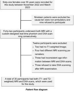
First, a real-time phantom consisting of oil and water tubes was manufactured. Then, 30 female volunteers (age: 62.3 ± 6.3 years) underwent lumbar spine examination with MRI (using a novel phantom) and dual-energy X-ray absorptiometry (DXA), following ethical approval. MRI phantom-based F-score and W-score were defined by normalizing the vertebral signal intensities (SIs) by the oil and water SIs of the phantom on T1- and T2-weighted images, respectively. The diagnostic performances of the new scores for assessing osteoporosis and vertebral fractures were examined using receiver operating characteristic analysis and compared with DXA-measured areal bone mineral density (DXA-aBMD).
Results
The F-score and W-score were greater in the osteoporotic patients (3.93 and 2.29) than the non-osteoporotic subjects (3.05 and 1.79) and achieved AUC values of 0.85 and 0.74 (p < 0.05), respectively, when detecting osteoporosis. Similarly, F-score and W-score had greater values for the fracture patients (3.94 and 2.53) than the non-fracture subjects (3.14 and 1.69) and produced better AUC values (0.90 for W-score and 0.79 for F-score) compared to DXA-aBMD (AUC: 0.27, p < 0.05). In addition, the F-score and W-score had a strong correlation (r = 0.77; p < 0.001).
Conclusion
A novel real-time lumber spine MRI phantom was developed, based upon which newly defined F-score and W-score were able to detect osteoporosis and demonstrated an improved ability over DXA-aBMD in differentiating patients with vertebral fractures.


 分享
分享

 求助内容:
求助内容: 应助结果提醒方式:
 扫码关注我们
