Dina Giese, Hao Li, Wei Liu, Karin Staxäng, Monika Hodik, Hanif M. Ladak, Sumit Agrawal, Anneliese Schrott-Fischer, Rudolf Glueckert, Helge Rask-Andersen
{"title":"人类科蒂隧道结构和耳蜗分区的显微解剖学--同位变异和跨细胞信号传导","authors":"Dina Giese, Hao Li, Wei Liu, Karin Staxäng, Monika Hodik, Hanif M. Ladak, Sumit Agrawal, Anneliese Schrott-Fischer, Rudolf Glueckert, Helge Rask-Andersen","doi":"10.1111/joa.14045","DOIUrl":null,"url":null,"abstract":"<p>Auditory sensitivity and frequency resolution depend on the optimal transfer of sound-induced vibrations from the basilar membrane (BM) to the inner hair cells (IHCs), the principal auditory receptors. There remains a paucity of information on how this is accomplished along the frequency range in the human cochlea. Most of the current knowledge is derived either from animal experiments or human tissue processed after death, offering limited structural preservation and optical resolution. In our study, we analyzed the cytoarchitecture of the human cochlear partition at different frequency locations using high-resolution microscopy of uniquely preserved normal human tissue. The results may have clinical implications and increase our understanding of how frequency-dependent acoustic vibrations are carried to human IHCs. A 1-micron-thick plastic-embedded section (mid-modiolar) from a normal human cochlea uniquely preserved at lateral skull base surgery was analyzed using light and transmission electron microscopy (LM, TEM). Frequency locations were estimated using synchrotron radiation phase-contrast imaging (SR-PCI). Archival human tissue prepared for scanning electron microscopy (SEM) and super-resolution structured illumination microscopy (SR-SIM) were also used and compared in this study. Microscopy demonstrated great variations in the dimension and architecture of the human cochlear partition along the frequency range. Pillar cell geometry was closely regulated and depended on the reticular lamina slope and tympanic lip angle. A type II collagen-expressing lamina extended medially from the tympanic lip under the inner sulcus, here named “accessory basilar membrane.” It was linked to the tympanic lip and inner pillar foot, and it may contribute to the overall compliance of the cochlear partition. Based on the findings, we speculate on the remarkable microanatomic inflections and geometric relationships which relay different sound-induced vibrations to the IHCs, including their relevance for the evolution of human speech reception and electric stimulation with auditory implants. The inner pillar transcellular microtubule/actin system's role of directly converting vibration energy to the IHC cuticular plate and ciliary bundle is highlighted.</p>","PeriodicalId":14971,"journal":{"name":"Journal of Anatomy","volume":null,"pages":null},"PeriodicalIF":1.8000,"publicationDate":"2024-04-13","publicationTypes":"Journal Article","fieldsOfStudy":null,"isOpenAccess":false,"openAccessPdf":"https://onlinelibrary.wiley.com/doi/epdf/10.1111/joa.14045","citationCount":"0","resultStr":"{\"title\":\"Microanatomy of the human tunnel of Corti structures and cochlear partition-tonotopic variations and transcellular signaling\",\"authors\":\"Dina Giese, Hao Li, Wei Liu, Karin Staxäng, Monika Hodik, Hanif M. Ladak, Sumit Agrawal, Anneliese Schrott-Fischer, Rudolf Glueckert, Helge Rask-Andersen\",\"doi\":\"10.1111/joa.14045\",\"DOIUrl\":null,\"url\":null,\"abstract\":\"<p>Auditory sensitivity and frequency resolution depend on the optimal transfer of sound-induced vibrations from the basilar membrane (BM) to the inner hair cells (IHCs), the principal auditory receptors. There remains a paucity of information on how this is accomplished along the frequency range in the human cochlea. Most of the current knowledge is derived either from animal experiments or human tissue processed after death, offering limited structural preservation and optical resolution. In our study, we analyzed the cytoarchitecture of the human cochlear partition at different frequency locations using high-resolution microscopy of uniquely preserved normal human tissue. The results may have clinical implications and increase our understanding of how frequency-dependent acoustic vibrations are carried to human IHCs. A 1-micron-thick plastic-embedded section (mid-modiolar) from a normal human cochlea uniquely preserved at lateral skull base surgery was analyzed using light and transmission electron microscopy (LM, TEM). Frequency locations were estimated using synchrotron radiation phase-contrast imaging (SR-PCI). Archival human tissue prepared for scanning electron microscopy (SEM) and super-resolution structured illumination microscopy (SR-SIM) were also used and compared in this study. Microscopy demonstrated great variations in the dimension and architecture of the human cochlear partition along the frequency range. Pillar cell geometry was closely regulated and depended on the reticular lamina slope and tympanic lip angle. A type II collagen-expressing lamina extended medially from the tympanic lip under the inner sulcus, here named “accessory basilar membrane.” It was linked to the tympanic lip and inner pillar foot, and it may contribute to the overall compliance of the cochlear partition. Based on the findings, we speculate on the remarkable microanatomic inflections and geometric relationships which relay different sound-induced vibrations to the IHCs, including their relevance for the evolution of human speech reception and electric stimulation with auditory implants. The inner pillar transcellular microtubule/actin system's role of directly converting vibration energy to the IHC cuticular plate and ciliary bundle is highlighted.</p>\",\"PeriodicalId\":14971,\"journal\":{\"name\":\"Journal of Anatomy\",\"volume\":null,\"pages\":null},\"PeriodicalIF\":1.8000,\"publicationDate\":\"2024-04-13\",\"publicationTypes\":\"Journal Article\",\"fieldsOfStudy\":null,\"isOpenAccess\":false,\"openAccessPdf\":\"https://onlinelibrary.wiley.com/doi/epdf/10.1111/joa.14045\",\"citationCount\":\"0\",\"resultStr\":null,\"platform\":\"Semanticscholar\",\"paperid\":null,\"PeriodicalName\":\"Journal of Anatomy\",\"FirstCategoryId\":\"3\",\"ListUrlMain\":\"https://onlinelibrary.wiley.com/doi/10.1111/joa.14045\",\"RegionNum\":3,\"RegionCategory\":\"医学\",\"ArticlePicture\":[],\"TitleCN\":null,\"AbstractTextCN\":null,\"PMCID\":null,\"EPubDate\":\"\",\"PubModel\":\"\",\"JCR\":\"Q2\",\"JCRName\":\"ANATOMY & MORPHOLOGY\",\"Score\":null,\"Total\":0}","platform":"Semanticscholar","paperid":null,"PeriodicalName":"Journal of Anatomy","FirstCategoryId":"3","ListUrlMain":"https://onlinelibrary.wiley.com/doi/10.1111/joa.14045","RegionNum":3,"RegionCategory":"医学","ArticlePicture":[],"TitleCN":null,"AbstractTextCN":null,"PMCID":null,"EPubDate":"","PubModel":"","JCR":"Q2","JCRName":"ANATOMY & MORPHOLOGY","Score":null,"Total":0}
Microanatomy of the human tunnel of Corti structures and cochlear partition-tonotopic variations and transcellular signaling
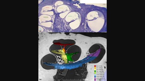
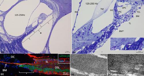
Auditory sensitivity and frequency resolution depend on the optimal transfer of sound-induced vibrations from the basilar membrane (BM) to the inner hair cells (IHCs), the principal auditory receptors. There remains a paucity of information on how this is accomplished along the frequency range in the human cochlea. Most of the current knowledge is derived either from animal experiments or human tissue processed after death, offering limited structural preservation and optical resolution. In our study, we analyzed the cytoarchitecture of the human cochlear partition at different frequency locations using high-resolution microscopy of uniquely preserved normal human tissue. The results may have clinical implications and increase our understanding of how frequency-dependent acoustic vibrations are carried to human IHCs. A 1-micron-thick plastic-embedded section (mid-modiolar) from a normal human cochlea uniquely preserved at lateral skull base surgery was analyzed using light and transmission electron microscopy (LM, TEM). Frequency locations were estimated using synchrotron radiation phase-contrast imaging (SR-PCI). Archival human tissue prepared for scanning electron microscopy (SEM) and super-resolution structured illumination microscopy (SR-SIM) were also used and compared in this study. Microscopy demonstrated great variations in the dimension and architecture of the human cochlear partition along the frequency range. Pillar cell geometry was closely regulated and depended on the reticular lamina slope and tympanic lip angle. A type II collagen-expressing lamina extended medially from the tympanic lip under the inner sulcus, here named “accessory basilar membrane.” It was linked to the tympanic lip and inner pillar foot, and it may contribute to the overall compliance of the cochlear partition. Based on the findings, we speculate on the remarkable microanatomic inflections and geometric relationships which relay different sound-induced vibrations to the IHCs, including their relevance for the evolution of human speech reception and electric stimulation with auditory implants. The inner pillar transcellular microtubule/actin system's role of directly converting vibration energy to the IHC cuticular plate and ciliary bundle is highlighted.
期刊介绍:
Journal of Anatomy is an international peer-reviewed journal sponsored by the Anatomical Society. The journal publishes original papers, invited review articles and book reviews. Its main focus is to understand anatomy through an analysis of structure, function, development and evolution. Priority will be given to studies of that clearly articulate their relevance to the anatomical community. Focal areas include: experimental studies, contributions based on molecular and cell biology and on the application of modern imaging techniques and papers with novel methods or synthetic perspective on an anatomical system.
Studies that are essentially descriptive anatomy are appropriate only if they communicate clearly a broader functional or evolutionary significance. You must clearly state the broader implications of your work in the abstract.
We particularly welcome submissions in the following areas:
Cell biology and tissue architecture
Comparative functional morphology
Developmental biology
Evolutionary developmental biology
Evolutionary morphology
Functional human anatomy
Integrative vertebrate paleontology
Methodological innovations in anatomical research
Musculoskeletal system
Neuroanatomy and neurodegeneration
Significant advances in anatomical education.


 分享
分享
 求助内容:
求助内容: 应助结果提醒方式:
 扫码关注我们
