Christoph Cremer, Florian Schock, Antonio Virgilio Failla, Udo Birk
{"title":"调制照明显微镜:核纳米结构分析的应用前景","authors":"Christoph Cremer, Florian Schock, Antonio Virgilio Failla, Udo Birk","doi":"10.1111/jmi.13297","DOIUrl":null,"url":null,"abstract":"<p>The structure of the cell nucleus of higher organisms has become a major topic of advanced light microscopy. So far, a variety of methods have been applied, including confocal laser scanning fluorescence microscopy, 4Pi, STED and localisation microscopy approaches, as well as different types of patterned illumination microscopy, modulated either laterally (in the object plane) or axially (along the optical axis). Based on our experience, we discuss here some application perspectives of Modulated Illumination Microscopy (MIM) and its combination with single-molecule localisation microscopy (SMLM). For example, spatially modulated illumination microscopy/SMI (illumination modulation along the optical axis) has been used to determine the axial extension (size) of small, optically isolated fluorescent objects between ≤ 200 nm and ≥ 40 nm diameter with a precision down to the few nm range; it also allows the axial positioning of such structures down to the 1 nm scale; combined with laterally structured illumination/SIM, a 3D localisation precision of ≤1 nm is expected using fluorescence yields typical for SMLM applications. Together with the nanosizing capability of SMI, this can be used to analyse macromolecular nuclear complexes with a resolution approaching that of cryoelectron microscopy.</p>","PeriodicalId":16484,"journal":{"name":"Journal of microscopy","volume":"296 2","pages":"121-128"},"PeriodicalIF":1.9000,"publicationDate":"2024-04-15","publicationTypes":"Journal Article","fieldsOfStudy":null,"isOpenAccess":false,"openAccessPdf":"https://onlinelibrary.wiley.com/doi/epdf/10.1111/jmi.13297","citationCount":"0","resultStr":"{\"title\":\"Modulated illumination microscopy: Application perspectives in nuclear nanostructure analysis\",\"authors\":\"Christoph Cremer, Florian Schock, Antonio Virgilio Failla, Udo Birk\",\"doi\":\"10.1111/jmi.13297\",\"DOIUrl\":null,\"url\":null,\"abstract\":\"<p>The structure of the cell nucleus of higher organisms has become a major topic of advanced light microscopy. So far, a variety of methods have been applied, including confocal laser scanning fluorescence microscopy, 4Pi, STED and localisation microscopy approaches, as well as different types of patterned illumination microscopy, modulated either laterally (in the object plane) or axially (along the optical axis). Based on our experience, we discuss here some application perspectives of Modulated Illumination Microscopy (MIM) and its combination with single-molecule localisation microscopy (SMLM). For example, spatially modulated illumination microscopy/SMI (illumination modulation along the optical axis) has been used to determine the axial extension (size) of small, optically isolated fluorescent objects between ≤ 200 nm and ≥ 40 nm diameter with a precision down to the few nm range; it also allows the axial positioning of such structures down to the 1 nm scale; combined with laterally structured illumination/SIM, a 3D localisation precision of ≤1 nm is expected using fluorescence yields typical for SMLM applications. Together with the nanosizing capability of SMI, this can be used to analyse macromolecular nuclear complexes with a resolution approaching that of cryoelectron microscopy.</p>\",\"PeriodicalId\":16484,\"journal\":{\"name\":\"Journal of microscopy\",\"volume\":\"296 2\",\"pages\":\"121-128\"},\"PeriodicalIF\":1.9000,\"publicationDate\":\"2024-04-15\",\"publicationTypes\":\"Journal Article\",\"fieldsOfStudy\":null,\"isOpenAccess\":false,\"openAccessPdf\":\"https://onlinelibrary.wiley.com/doi/epdf/10.1111/jmi.13297\",\"citationCount\":\"0\",\"resultStr\":null,\"platform\":\"Semanticscholar\",\"paperid\":null,\"PeriodicalName\":\"Journal of microscopy\",\"FirstCategoryId\":\"5\",\"ListUrlMain\":\"https://onlinelibrary.wiley.com/doi/10.1111/jmi.13297\",\"RegionNum\":4,\"RegionCategory\":\"工程技术\",\"ArticlePicture\":[],\"TitleCN\":null,\"AbstractTextCN\":null,\"PMCID\":null,\"EPubDate\":\"\",\"PubModel\":\"\",\"JCR\":\"Q3\",\"JCRName\":\"MICROSCOPY\",\"Score\":null,\"Total\":0}","platform":"Semanticscholar","paperid":null,"PeriodicalName":"Journal of microscopy","FirstCategoryId":"5","ListUrlMain":"https://onlinelibrary.wiley.com/doi/10.1111/jmi.13297","RegionNum":4,"RegionCategory":"工程技术","ArticlePicture":[],"TitleCN":null,"AbstractTextCN":null,"PMCID":null,"EPubDate":"","PubModel":"","JCR":"Q3","JCRName":"MICROSCOPY","Score":null,"Total":0}
Modulated illumination microscopy: Application perspectives in nuclear nanostructure analysis
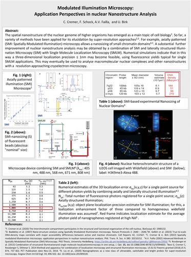
The structure of the cell nucleus of higher organisms has become a major topic of advanced light microscopy. So far, a variety of methods have been applied, including confocal laser scanning fluorescence microscopy, 4Pi, STED and localisation microscopy approaches, as well as different types of patterned illumination microscopy, modulated either laterally (in the object plane) or axially (along the optical axis). Based on our experience, we discuss here some application perspectives of Modulated Illumination Microscopy (MIM) and its combination with single-molecule localisation microscopy (SMLM). For example, spatially modulated illumination microscopy/SMI (illumination modulation along the optical axis) has been used to determine the axial extension (size) of small, optically isolated fluorescent objects between ≤ 200 nm and ≥ 40 nm diameter with a precision down to the few nm range; it also allows the axial positioning of such structures down to the 1 nm scale; combined with laterally structured illumination/SIM, a 3D localisation precision of ≤1 nm is expected using fluorescence yields typical for SMLM applications. Together with the nanosizing capability of SMI, this can be used to analyse macromolecular nuclear complexes with a resolution approaching that of cryoelectron microscopy.
期刊介绍:
The Journal of Microscopy is the oldest journal dedicated to the science of microscopy and the only peer-reviewed publication of the Royal Microscopical Society. It publishes papers that report on the very latest developments in microscopy such as advances in microscopy techniques or novel areas of application. The Journal does not seek to publish routine applications of microscopy or specimen preparation even though the submission may otherwise have a high scientific merit.
The scope covers research in the physical and biological sciences and covers imaging methods using light, electrons, X-rays and other radiations as well as atomic force and near field techniques. Interdisciplinary research is welcome. Papers pertaining to microscopy are also welcomed on optical theory, spectroscopy, novel specimen preparation and manipulation methods and image recording, processing and analysis including dynamic analysis of living specimens.
Publication types include full papers, hot topic fast tracked communications and review articles. Authors considering submitting a review article should contact the editorial office first.


 分享
分享
 求助内容:
求助内容: 应助结果提醒方式:
 扫码关注我们
