Ronaldo Roberto Tait Caleffe , Stefany Rodrigues de Oliveira , Eduardo Amat , Luz Miryam Gomez-Piñerez , Bruno Vinicius Daquila , Maria Claudia Colla Ruvulo-Takasusuki , Helio Conte
{"title":"传入的蝇类 Chrysomya megacephala (Fabricius, 1794) 的幼虫内部形态(双翅目:茧蝇科)","authors":"Ronaldo Roberto Tait Caleffe , Stefany Rodrigues de Oliveira , Eduardo Amat , Luz Miryam Gomez-Piñerez , Bruno Vinicius Daquila , Maria Claudia Colla Ruvulo-Takasusuki , Helio Conte","doi":"10.1016/j.jcz.2024.03.003","DOIUrl":null,"url":null,"abstract":"<div><p>The blowfly <em>Chrysomya megacephala</em> (Diptera: Calliphoridae) colonizes different environments and is found in many global regions. Its importance includes its involvement in the decomposition of organic matter, forensic entomology, maggot therapy, bioprospecting, source of biodiesel, industry, stabilization of heavy metals in soil, pollination, and as a vector of diseases and causing secondary myiasis. Internal morphological studies contribute to understanding insect's function and provide basic data for applied areas with interest in this specie. The objective of this study was to describe the internal morphology of <em>C. megacephala</em> larvae. Post-embryonic development occurred over 240 h, equally divided into larval and pupal stages. After hatching, the larvae averaged <1 mm in length and 0.02 mg in weight, growing to 13.83 mm and 50.4 mg in the third instar. We utilized whole, third-instar larvae to perform histology. Sensory organs were observed in the cephalic region, antennae, maxillary palps, and ventral organ. The alimentary canal comprises a foregut, midgut, and hindgut, including tracheal insertions and accessory organs, such as salivary glands. The midgut is the largest organ of the alimentary canal and fat body are present in body cavity and intersegmental space. Thus, our data about the internal morphology of <em>C. megacephala</em> larvae describe the structure and position of the alimentary canal, salivary gland, sensory organs, and fat body.</p></div>","PeriodicalId":49332,"journal":{"name":"Zoologischer Anzeiger","volume":"310 ","pages":"Pages 23-33"},"PeriodicalIF":1.2000,"publicationDate":"2024-04-02","publicationTypes":"Journal Article","fieldsOfStudy":null,"isOpenAccess":false,"openAccessPdf":"","citationCount":"0","resultStr":"{\"title\":\"Larval internal morphology of the introduced blowfly Chrysomya megacephala (Fabricius, 1794) (Diptera: Calliphoridae)\",\"authors\":\"Ronaldo Roberto Tait Caleffe , Stefany Rodrigues de Oliveira , Eduardo Amat , Luz Miryam Gomez-Piñerez , Bruno Vinicius Daquila , Maria Claudia Colla Ruvulo-Takasusuki , Helio Conte\",\"doi\":\"10.1016/j.jcz.2024.03.003\",\"DOIUrl\":null,\"url\":null,\"abstract\":\"<div><p>The blowfly <em>Chrysomya megacephala</em> (Diptera: Calliphoridae) colonizes different environments and is found in many global regions. Its importance includes its involvement in the decomposition of organic matter, forensic entomology, maggot therapy, bioprospecting, source of biodiesel, industry, stabilization of heavy metals in soil, pollination, and as a vector of diseases and causing secondary myiasis. Internal morphological studies contribute to understanding insect's function and provide basic data for applied areas with interest in this specie. The objective of this study was to describe the internal morphology of <em>C. megacephala</em> larvae. Post-embryonic development occurred over 240 h, equally divided into larval and pupal stages. After hatching, the larvae averaged <1 mm in length and 0.02 mg in weight, growing to 13.83 mm and 50.4 mg in the third instar. We utilized whole, third-instar larvae to perform histology. Sensory organs were observed in the cephalic region, antennae, maxillary palps, and ventral organ. The alimentary canal comprises a foregut, midgut, and hindgut, including tracheal insertions and accessory organs, such as salivary glands. The midgut is the largest organ of the alimentary canal and fat body are present in body cavity and intersegmental space. Thus, our data about the internal morphology of <em>C. megacephala</em> larvae describe the structure and position of the alimentary canal, salivary gland, sensory organs, and fat body.</p></div>\",\"PeriodicalId\":49332,\"journal\":{\"name\":\"Zoologischer Anzeiger\",\"volume\":\"310 \",\"pages\":\"Pages 23-33\"},\"PeriodicalIF\":1.2000,\"publicationDate\":\"2024-04-02\",\"publicationTypes\":\"Journal Article\",\"fieldsOfStudy\":null,\"isOpenAccess\":false,\"openAccessPdf\":\"\",\"citationCount\":\"0\",\"resultStr\":null,\"platform\":\"Semanticscholar\",\"paperid\":null,\"PeriodicalName\":\"Zoologischer Anzeiger\",\"FirstCategoryId\":\"99\",\"ListUrlMain\":\"https://www.sciencedirect.com/science/article/pii/S0044523124000275\",\"RegionNum\":3,\"RegionCategory\":\"生物学\",\"ArticlePicture\":[],\"TitleCN\":null,\"AbstractTextCN\":null,\"PMCID\":null,\"EPubDate\":\"\",\"PubModel\":\"\",\"JCR\":\"Q2\",\"JCRName\":\"ZOOLOGY\",\"Score\":null,\"Total\":0}","platform":"Semanticscholar","paperid":null,"PeriodicalName":"Zoologischer Anzeiger","FirstCategoryId":"99","ListUrlMain":"https://www.sciencedirect.com/science/article/pii/S0044523124000275","RegionNum":3,"RegionCategory":"生物学","ArticlePicture":[],"TitleCN":null,"AbstractTextCN":null,"PMCID":null,"EPubDate":"","PubModel":"","JCR":"Q2","JCRName":"ZOOLOGY","Score":null,"Total":0}
Larval internal morphology of the introduced blowfly Chrysomya megacephala (Fabricius, 1794) (Diptera: Calliphoridae)
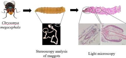
The blowfly Chrysomya megacephala (Diptera: Calliphoridae) colonizes different environments and is found in many global regions. Its importance includes its involvement in the decomposition of organic matter, forensic entomology, maggot therapy, bioprospecting, source of biodiesel, industry, stabilization of heavy metals in soil, pollination, and as a vector of diseases and causing secondary myiasis. Internal morphological studies contribute to understanding insect's function and provide basic data for applied areas with interest in this specie. The objective of this study was to describe the internal morphology of C. megacephala larvae. Post-embryonic development occurred over 240 h, equally divided into larval and pupal stages. After hatching, the larvae averaged <1 mm in length and 0.02 mg in weight, growing to 13.83 mm and 50.4 mg in the third instar. We utilized whole, third-instar larvae to perform histology. Sensory organs were observed in the cephalic region, antennae, maxillary palps, and ventral organ. The alimentary canal comprises a foregut, midgut, and hindgut, including tracheal insertions and accessory organs, such as salivary glands. The midgut is the largest organ of the alimentary canal and fat body are present in body cavity and intersegmental space. Thus, our data about the internal morphology of C. megacephala larvae describe the structure and position of the alimentary canal, salivary gland, sensory organs, and fat body.
期刊介绍:
Zoologischer Anzeiger - A Journal of Comparative Zoology is devoted to comparative zoology with a special emphasis on morphology, systematics, biogeography, and evolutionary biology targeting all metazoans, both modern and extinct. We also consider taxonomic submissions addressing a broader systematic and/or evolutionary context. The overall aim of the journal is to contribute to our understanding of the organismic world from an evolutionary perspective.
The journal Zoologischer Anzeiger invites suggestions for special issues. Interested parties may contact one of the editors.


 分享
分享
 求助内容:
求助内容: 应助结果提醒方式:
 扫码关注我们
