{"title":"伴有或不伴有腭裂的齿槽裂患者的全景外观差异。","authors":"Takeshi Fujii, Chiaki Kuwada, Yoshitaka Kise, Motoki Fukuda, Mizuho Mori, Masako Nishiyama, Michihito Nozawa, Munetaka Naitoh, Yoshiko Ariji, Eiichiro Ariji","doi":"10.5624/isd.20230159","DOIUrl":null,"url":null,"abstract":"<p><strong>Purpose: </strong>The purpose of this study was to clarify the panoramic image differences of cleft alveolus patients with or without a cleft palate, with emphases on the visibility of the line formed by the junction between the nasal septum and nasal floor (the upper line) and the appearances of the maxillary lateral incisor.</p><p><strong>Materials and methods: </strong>Panoramic radiographs of 238 patients with cleft alveolus were analyzed for the visibility of the upper line, including clear, obscure or invisible, and the appearances of the maxillary lateral incisor, regarding congenital absence, incomplete growth, delayed eruption and medial inclination. Differences in the distribution ratio of these visibility and appearances were verified between the patients with and without a cleft palate using the chi-square test.</p><p><strong>Results: </strong>There was a significant difference in the visibility distribution of the upper line between the patients with and without a cleft palate (p<0.05). In most of the patients with a cleft palate, the upper line was not observed. In the unilateral cleft alveolus patients, the medial inclination of the maxillary lateral incisor was more frequently observed in patients with a cleft palate than in patients without a cleft palate.</p><p><strong>Conclusion: </strong>Two differences were identified in panoramic appearances. The first was the disappearance (invisible appearance) of the upper line in patients with a cleft palate, and the second was a change in the medial inclination on the affected side maxillary lateral incisor in unilateral cleft alveolus patients with a cleft palate.</p>","PeriodicalId":51714,"journal":{"name":"Imaging Science in Dentistry","volume":"54 1","pages":"25-31"},"PeriodicalIF":2.1000,"publicationDate":"2024-03-01","publicationTypes":"Journal Article","fieldsOfStudy":null,"isOpenAccess":false,"openAccessPdf":"https://www.ncbi.nlm.nih.gov/pmc/articles/PMC10985517/pdf/","citationCount":"0","resultStr":"{\"title\":\"Differences in the panoramic appearance of cleft alveolus patients with or without a cleft palate.\",\"authors\":\"Takeshi Fujii, Chiaki Kuwada, Yoshitaka Kise, Motoki Fukuda, Mizuho Mori, Masako Nishiyama, Michihito Nozawa, Munetaka Naitoh, Yoshiko Ariji, Eiichiro Ariji\",\"doi\":\"10.5624/isd.20230159\",\"DOIUrl\":null,\"url\":null,\"abstract\":\"<p><strong>Purpose: </strong>The purpose of this study was to clarify the panoramic image differences of cleft alveolus patients with or without a cleft palate, with emphases on the visibility of the line formed by the junction between the nasal septum and nasal floor (the upper line) and the appearances of the maxillary lateral incisor.</p><p><strong>Materials and methods: </strong>Panoramic radiographs of 238 patients with cleft alveolus were analyzed for the visibility of the upper line, including clear, obscure or invisible, and the appearances of the maxillary lateral incisor, regarding congenital absence, incomplete growth, delayed eruption and medial inclination. Differences in the distribution ratio of these visibility and appearances were verified between the patients with and without a cleft palate using the chi-square test.</p><p><strong>Results: </strong>There was a significant difference in the visibility distribution of the upper line between the patients with and without a cleft palate (p<0.05). In most of the patients with a cleft palate, the upper line was not observed. In the unilateral cleft alveolus patients, the medial inclination of the maxillary lateral incisor was more frequently observed in patients with a cleft palate than in patients without a cleft palate.</p><p><strong>Conclusion: </strong>Two differences were identified in panoramic appearances. The first was the disappearance (invisible appearance) of the upper line in patients with a cleft palate, and the second was a change in the medial inclination on the affected side maxillary lateral incisor in unilateral cleft alveolus patients with a cleft palate.</p>\",\"PeriodicalId\":51714,\"journal\":{\"name\":\"Imaging Science in Dentistry\",\"volume\":\"54 1\",\"pages\":\"25-31\"},\"PeriodicalIF\":2.1000,\"publicationDate\":\"2024-03-01\",\"publicationTypes\":\"Journal Article\",\"fieldsOfStudy\":null,\"isOpenAccess\":false,\"openAccessPdf\":\"https://www.ncbi.nlm.nih.gov/pmc/articles/PMC10985517/pdf/\",\"citationCount\":\"0\",\"resultStr\":null,\"platform\":\"Semanticscholar\",\"paperid\":null,\"PeriodicalName\":\"Imaging Science in Dentistry\",\"FirstCategoryId\":\"1085\",\"ListUrlMain\":\"https://doi.org/10.5624/isd.20230159\",\"RegionNum\":0,\"RegionCategory\":null,\"ArticlePicture\":[],\"TitleCN\":null,\"AbstractTextCN\":null,\"PMCID\":null,\"EPubDate\":\"2023/12/13 0:00:00\",\"PubModel\":\"Epub\",\"JCR\":\"Q3\",\"JCRName\":\"DENTISTRY, ORAL SURGERY & MEDICINE\",\"Score\":null,\"Total\":0}","platform":"Semanticscholar","paperid":null,"PeriodicalName":"Imaging Science in Dentistry","FirstCategoryId":"1085","ListUrlMain":"https://doi.org/10.5624/isd.20230159","RegionNum":0,"RegionCategory":null,"ArticlePicture":[],"TitleCN":null,"AbstractTextCN":null,"PMCID":null,"EPubDate":"2023/12/13 0:00:00","PubModel":"Epub","JCR":"Q3","JCRName":"DENTISTRY, ORAL SURGERY & MEDICINE","Score":null,"Total":0}
Differences in the panoramic appearance of cleft alveolus patients with or without a cleft palate.
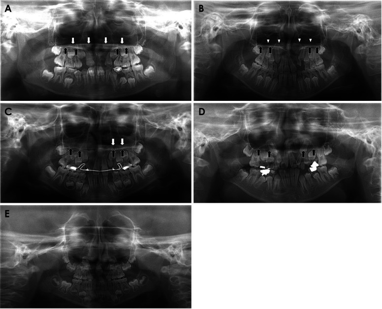
Purpose: The purpose of this study was to clarify the panoramic image differences of cleft alveolus patients with or without a cleft palate, with emphases on the visibility of the line formed by the junction between the nasal septum and nasal floor (the upper line) and the appearances of the maxillary lateral incisor.
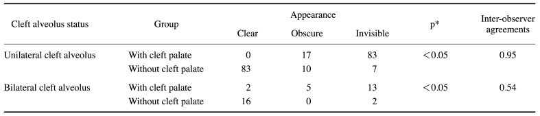
Materials and methods: Panoramic radiographs of 238 patients with cleft alveolus were analyzed for the visibility of the upper line, including clear, obscure or invisible, and the appearances of the maxillary lateral incisor, regarding congenital absence, incomplete growth, delayed eruption and medial inclination. Differences in the distribution ratio of these visibility and appearances were verified between the patients with and without a cleft palate using the chi-square test.
Results: There was a significant difference in the visibility distribution of the upper line between the patients with and without a cleft palate (p<0.05). In most of the patients with a cleft palate, the upper line was not observed. In the unilateral cleft alveolus patients, the medial inclination of the maxillary lateral incisor was more frequently observed in patients with a cleft palate than in patients without a cleft palate.
Conclusion: Two differences were identified in panoramic appearances. The first was the disappearance (invisible appearance) of the upper line in patients with a cleft palate, and the second was a change in the medial inclination on the affected side maxillary lateral incisor in unilateral cleft alveolus patients with a cleft palate.


 分享
分享
 求助内容:
求助内容: 应助结果提醒方式:
 扫码关注我们
