Shanti Joseph , Elizabeth Cherian , M.T. Ramesan , Sarath G. Nair , Reedhu Raj
{"title":"体内合成氧化锌纳米粒子的生物学评价和分子模型研究","authors":"Shanti Joseph , Elizabeth Cherian , M.T. Ramesan , Sarath G. Nair , Reedhu Raj","doi":"10.1016/j.nanoso.2024.101172","DOIUrl":null,"url":null,"abstract":"<div><p>The ZnO nanoparticle is an excellent candidate for biological applications. It has potential antimicrobial, antioxidant, anticancerous, antidiabetic, anti-inflammatory and wound healing properties because of its biodegradability and biocompatibility. The green method of synthesizing nanoparticles is gaining popularity as it is cost effective and reduces the impact of toxic substances. In the present work, <em>Eichhornia crassipes</em> was used as the plant source for the synthesis of ZnO nanoparticles. It is an invasive aquatic macrophyte that is utilized to exploit its phytoaccumulation property. The goal of the current study was to evaluate the ability of a living plant to transform the accumulated metal into metal nanoparticles <em>in vivo</em>. The formation of ZnO nanoparticles was confirmed by UV–visible spectrophotometry, EDX analysis, FTIR, XRD and HRTEM. A strong absorption peak (300 nm) and an excitonic peak (243 nm) obtained in the UV spectrophotometric analysis confirmed the formation of ZnO nanoparticles. The presence of strong signals for zinc and oxygen in the extracted nanoparticles was identified by EDX analysis. The presence of proteins, alkaloids, flavonoids and phenolics were identified using FTIR and are contributed to the formation of ZnO nps by the reduction reaction. XRD analysis revealed the hexagonal phase wurtzite structure of ZnO with a crystalline size of 16.89 nm. HRTEM analysis revealed that the particles were spherical and agglomerated in nature with an average size of 16 nm which is consistent with the XRD results. The ZnO nanoparticles were evaluated for their antibacterial activity against pathogenic bacterial strains such as <em>Escherichia coli, Pseudomonas aeruginosa, Staphylococcus aureus</em> and <em>Klebsiella pneumoniae.</em> The antibacterial activity of the ZnO nanoparticles was found to increase with their increasing concentration. The anticancerous activity of ZnO nanoparticles was also evaluated and exhibited a dose-dependent cytotoxicity against MCF-7 cells, which was further confirmed with molecular docking studies. Overall, a rapid, economical and ecofriendly approach for extracting ZnO nanoparticles was established, which can be employed as a potential therapeutic agent, particularly in nanomedicine for bacterial and cancer treatment.</p></div>","PeriodicalId":397,"journal":{"name":"Nano-Structures & Nano-Objects","volume":"38 ","pages":"Article 101172"},"PeriodicalIF":5.4500,"publicationDate":"2024-05-01","publicationTypes":"Journal Article","fieldsOfStudy":null,"isOpenAccess":false,"openAccessPdf":"","citationCount":"0","resultStr":"{\"title\":\"Biological evaluation and molecular modelling studies of in vivo synthesized ZnO nanoparticles\",\"authors\":\"Shanti Joseph , Elizabeth Cherian , M.T. Ramesan , Sarath G. Nair , Reedhu Raj\",\"doi\":\"10.1016/j.nanoso.2024.101172\",\"DOIUrl\":null,\"url\":null,\"abstract\":\"<div><p>The ZnO nanoparticle is an excellent candidate for biological applications. It has potential antimicrobial, antioxidant, anticancerous, antidiabetic, anti-inflammatory and wound healing properties because of its biodegradability and biocompatibility. The green method of synthesizing nanoparticles is gaining popularity as it is cost effective and reduces the impact of toxic substances. In the present work, <em>Eichhornia crassipes</em> was used as the plant source for the synthesis of ZnO nanoparticles. It is an invasive aquatic macrophyte that is utilized to exploit its phytoaccumulation property. The goal of the current study was to evaluate the ability of a living plant to transform the accumulated metal into metal nanoparticles <em>in vivo</em>. The formation of ZnO nanoparticles was confirmed by UV–visible spectrophotometry, EDX analysis, FTIR, XRD and HRTEM. A strong absorption peak (300 nm) and an excitonic peak (243 nm) obtained in the UV spectrophotometric analysis confirmed the formation of ZnO nanoparticles. The presence of strong signals for zinc and oxygen in the extracted nanoparticles was identified by EDX analysis. The presence of proteins, alkaloids, flavonoids and phenolics were identified using FTIR and are contributed to the formation of ZnO nps by the reduction reaction. XRD analysis revealed the hexagonal phase wurtzite structure of ZnO with a crystalline size of 16.89 nm. HRTEM analysis revealed that the particles were spherical and agglomerated in nature with an average size of 16 nm which is consistent with the XRD results. The ZnO nanoparticles were evaluated for their antibacterial activity against pathogenic bacterial strains such as <em>Escherichia coli, Pseudomonas aeruginosa, Staphylococcus aureus</em> and <em>Klebsiella pneumoniae.</em> The antibacterial activity of the ZnO nanoparticles was found to increase with their increasing concentration. The anticancerous activity of ZnO nanoparticles was also evaluated and exhibited a dose-dependent cytotoxicity against MCF-7 cells, which was further confirmed with molecular docking studies. Overall, a rapid, economical and ecofriendly approach for extracting ZnO nanoparticles was established, which can be employed as a potential therapeutic agent, particularly in nanomedicine for bacterial and cancer treatment.</p></div>\",\"PeriodicalId\":397,\"journal\":{\"name\":\"Nano-Structures & Nano-Objects\",\"volume\":\"38 \",\"pages\":\"Article 101172\"},\"PeriodicalIF\":5.4500,\"publicationDate\":\"2024-05-01\",\"publicationTypes\":\"Journal Article\",\"fieldsOfStudy\":null,\"isOpenAccess\":false,\"openAccessPdf\":\"\",\"citationCount\":\"0\",\"resultStr\":null,\"platform\":\"Semanticscholar\",\"paperid\":null,\"PeriodicalName\":\"Nano-Structures & Nano-Objects\",\"FirstCategoryId\":\"1\",\"ListUrlMain\":\"https://www.sciencedirect.com/science/article/pii/S2352507X24000830\",\"RegionNum\":0,\"RegionCategory\":null,\"ArticlePicture\":[],\"TitleCN\":null,\"AbstractTextCN\":null,\"PMCID\":null,\"EPubDate\":\"2024/5/8 0:00:00\",\"PubModel\":\"Epub\",\"JCR\":\"Q1\",\"JCRName\":\"Physics and Astronomy\",\"Score\":null,\"Total\":0}","platform":"Semanticscholar","paperid":null,"PeriodicalName":"Nano-Structures & Nano-Objects","FirstCategoryId":"1","ListUrlMain":"https://www.sciencedirect.com/science/article/pii/S2352507X24000830","RegionNum":0,"RegionCategory":null,"ArticlePicture":[],"TitleCN":null,"AbstractTextCN":null,"PMCID":null,"EPubDate":"2024/5/8 0:00:00","PubModel":"Epub","JCR":"Q1","JCRName":"Physics and Astronomy","Score":null,"Total":0}
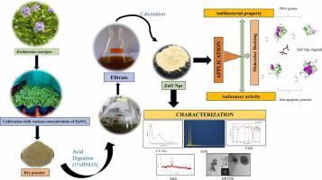
Biological evaluation and molecular modelling studies of in vivo synthesized ZnO nanoparticles
The ZnO nanoparticle is an excellent candidate for biological applications. It has potential antimicrobial, antioxidant, anticancerous, antidiabetic, anti-inflammatory and wound healing properties because of its biodegradability and biocompatibility. The green method of synthesizing nanoparticles is gaining popularity as it is cost effective and reduces the impact of toxic substances. In the present work, Eichhornia crassipes was used as the plant source for the synthesis of ZnO nanoparticles. It is an invasive aquatic macrophyte that is utilized to exploit its phytoaccumulation property. The goal of the current study was to evaluate the ability of a living plant to transform the accumulated metal into metal nanoparticles in vivo. The formation of ZnO nanoparticles was confirmed by UV–visible spectrophotometry, EDX analysis, FTIR, XRD and HRTEM. A strong absorption peak (300 nm) and an excitonic peak (243 nm) obtained in the UV spectrophotometric analysis confirmed the formation of ZnO nanoparticles. The presence of strong signals for zinc and oxygen in the extracted nanoparticles was identified by EDX analysis. The presence of proteins, alkaloids, flavonoids and phenolics were identified using FTIR and are contributed to the formation of ZnO nps by the reduction reaction. XRD analysis revealed the hexagonal phase wurtzite structure of ZnO with a crystalline size of 16.89 nm. HRTEM analysis revealed that the particles were spherical and agglomerated in nature with an average size of 16 nm which is consistent with the XRD results. The ZnO nanoparticles were evaluated for their antibacterial activity against pathogenic bacterial strains such as Escherichia coli, Pseudomonas aeruginosa, Staphylococcus aureus and Klebsiella pneumoniae. The antibacterial activity of the ZnO nanoparticles was found to increase with their increasing concentration. The anticancerous activity of ZnO nanoparticles was also evaluated and exhibited a dose-dependent cytotoxicity against MCF-7 cells, which was further confirmed with molecular docking studies. Overall, a rapid, economical and ecofriendly approach for extracting ZnO nanoparticles was established, which can be employed as a potential therapeutic agent, particularly in nanomedicine for bacterial and cancer treatment.
期刊介绍:
Nano-Structures & Nano-Objects is a new journal devoted to all aspects of the synthesis and the properties of this new flourishing domain. The journal is devoted to novel architectures at the nano-level with an emphasis on new synthesis and characterization methods. The journal is focused on the objects rather than on their applications. However, the research for new applications of original nano-structures & nano-objects in various fields such as nano-electronics, energy conversion, catalysis, drug delivery and nano-medicine is also welcome. The scope of Nano-Structures & Nano-Objects involves: -Metal and alloy nanoparticles with complex nanostructures such as shape control, core-shell and dumbells -Oxide nanoparticles and nanostructures, with complex oxide/metal, oxide/surface and oxide /organic interfaces -Inorganic semi-conducting nanoparticles (quantum dots) with an emphasis on new phases, structures, shapes and complexity -Nanostructures involving molecular inorganic species such as nanoparticles of coordination compounds, molecular magnets, spin transition nanoparticles etc. or organic nano-objects, in particular for molecular electronics -Nanostructured materials such as nano-MOFs and nano-zeolites -Hetero-junctions between molecules and nano-objects, between different nano-objects & nanostructures or between nano-objects & nanostructures and surfaces -Methods of characterization specific of the nano size or adapted for the nano size such as X-ray and neutron scattering, light scattering, NMR, Raman, Plasmonics, near field microscopies, various TEM and SEM techniques, magnetic studies, etc .


 分享
分享
 求助内容:
求助内容: 应助结果提醒方式:
 扫码关注我们
