{"title":"利用人体胚胎干细胞衍生的视网膜色素上皮细胞进行布鲁氏膜再生的电纺聚左旋乳酸膜的细胞相容性。","authors":"Naghmeh Abbasi, Helen O'Neill","doi":"10.1002/jbm.a.37736","DOIUrl":null,"url":null,"abstract":"<p>Cell replacement therapy is under development for dry age-related macular degeneration (AMD). A thin membrane resembling the Bruch's membrane is required to form a cell-on-membrane construct with retinal pigment epithelial (RPE) cells. These cells have been differentiated from human embryonic stem cells (hESCs) <i>in vitro</i>. A carrier membrane is required for cell implantation, which is biocompatible for cell growth and has dimensions and physical properties resembling the Bruch's membrane. Here a nanofiber electrospun poly-L-lactic acid (PLLA) membrane is tested for capacity to support cell growth and maturation. The requirements for laminin coating of the membrane are identified here. A porous electrospun nanofibrous PLLA membrane of ∼50 nm fiber diameter was developed as a prototype support for functional RPE cells grown as a monolayer. The need for laminin coating applied to the membrane following treatment with poly-L-ornithine (PLO), was identified in terms of cell growth and survival. Test membranes were compared in terms of hydrophilicity after laminin coating, mechanical properties of surface roughness and Young's modulus, porosity and ability to promote the attachment and proliferation of hESC-RPE cells in culture for up to 8 weeks. Over this time, RPE cell proliferation, morphology, and marker and gene expression, were monitored. The functional capacity of cell monolayers was identified in terms of transepithelial electrical resistance (TEER), phagocytosis of cells, as well as expression of the cytokines, vascular endothelial growth factor (VEGF) and pigment epithelium-derived factor (PEDF). PLLA polymer fibers are naturally hydrophobic, so their hydrophilicity was improved by pretreatment with PLO for subsequent coating with the bioactive protein laminin. They were then assessed for amount of laminin adsorbed, contact angle and uniformity of coating using scanning electron microscopy (SEM). Pretreatment with 100% PLO gave the best result over 10% PLO treatment or no treatment prior to laminin adsorption with significantly greater surface stiffness and modulus. By 6 weeks after cell plating, the coated membranes could support a mature RPE monolayer showing a dense apical microvillus structure and pigmented 3D polygonal cell morphology. After 8 weeks, PLO (100%)-Lam coated membranes exhibited the highest cell number, cell proliferation, and RPE barrier function measured as TEER. RPE cells showed the higher levels of specific surface marker and gene expression. Microphthalmia-associated transcription factor expression was highly upregulated indicating maturation of cells. Functionality of cells was indicated by expression of <i>VEGF</i> and <i>PEDF</i> genes as well as phagocytic capacity. In conclusion, electrospun PLLA membranes coated with PLO-Lam have the physical and biological properties to support the distribution and migration of hESC-RPE cells throughout the whole structure. They represent a good membrane candidate for preparation of hESC-RPE cells as a monolayer for implantation into the subretinal space of AMD patients.</p>","PeriodicalId":15142,"journal":{"name":"Journal of biomedical materials research. Part A","volume":"112 11","pages":"1902-1920"},"PeriodicalIF":3.9000,"publicationDate":"2024-05-10","publicationTypes":"Journal Article","fieldsOfStudy":null,"isOpenAccess":false,"openAccessPdf":"https://onlinelibrary.wiley.com/doi/epdf/10.1002/jbm.a.37736","citationCount":"0","resultStr":"{\"title\":\"Cytocompatibility of electrospun poly-L-lactic acid membranes for Bruch's membrane regeneration using human embryonic stem cell-derived retinal pigment epithelial cells\",\"authors\":\"Naghmeh Abbasi, Helen O'Neill\",\"doi\":\"10.1002/jbm.a.37736\",\"DOIUrl\":null,\"url\":null,\"abstract\":\"<p>Cell replacement therapy is under development for dry age-related macular degeneration (AMD). A thin membrane resembling the Bruch's membrane is required to form a cell-on-membrane construct with retinal pigment epithelial (RPE) cells. These cells have been differentiated from human embryonic stem cells (hESCs) <i>in vitro</i>. A carrier membrane is required for cell implantation, which is biocompatible for cell growth and has dimensions and physical properties resembling the Bruch's membrane. Here a nanofiber electrospun poly-L-lactic acid (PLLA) membrane is tested for capacity to support cell growth and maturation. The requirements for laminin coating of the membrane are identified here. A porous electrospun nanofibrous PLLA membrane of ∼50 nm fiber diameter was developed as a prototype support for functional RPE cells grown as a monolayer. The need for laminin coating applied to the membrane following treatment with poly-L-ornithine (PLO), was identified in terms of cell growth and survival. Test membranes were compared in terms of hydrophilicity after laminin coating, mechanical properties of surface roughness and Young's modulus, porosity and ability to promote the attachment and proliferation of hESC-RPE cells in culture for up to 8 weeks. Over this time, RPE cell proliferation, morphology, and marker and gene expression, were monitored. The functional capacity of cell monolayers was identified in terms of transepithelial electrical resistance (TEER), phagocytosis of cells, as well as expression of the cytokines, vascular endothelial growth factor (VEGF) and pigment epithelium-derived factor (PEDF). PLLA polymer fibers are naturally hydrophobic, so their hydrophilicity was improved by pretreatment with PLO for subsequent coating with the bioactive protein laminin. They were then assessed for amount of laminin adsorbed, contact angle and uniformity of coating using scanning electron microscopy (SEM). Pretreatment with 100% PLO gave the best result over 10% PLO treatment or no treatment prior to laminin adsorption with significantly greater surface stiffness and modulus. By 6 weeks after cell plating, the coated membranes could support a mature RPE monolayer showing a dense apical microvillus structure and pigmented 3D polygonal cell morphology. After 8 weeks, PLO (100%)-Lam coated membranes exhibited the highest cell number, cell proliferation, and RPE barrier function measured as TEER. RPE cells showed the higher levels of specific surface marker and gene expression. Microphthalmia-associated transcription factor expression was highly upregulated indicating maturation of cells. Functionality of cells was indicated by expression of <i>VEGF</i> and <i>PEDF</i> genes as well as phagocytic capacity. In conclusion, electrospun PLLA membranes coated with PLO-Lam have the physical and biological properties to support the distribution and migration of hESC-RPE cells throughout the whole structure. They represent a good membrane candidate for preparation of hESC-RPE cells as a monolayer for implantation into the subretinal space of AMD patients.</p>\",\"PeriodicalId\":15142,\"journal\":{\"name\":\"Journal of biomedical materials research. Part A\",\"volume\":\"112 11\",\"pages\":\"1902-1920\"},\"PeriodicalIF\":3.9000,\"publicationDate\":\"2024-05-10\",\"publicationTypes\":\"Journal Article\",\"fieldsOfStudy\":null,\"isOpenAccess\":false,\"openAccessPdf\":\"https://onlinelibrary.wiley.com/doi/epdf/10.1002/jbm.a.37736\",\"citationCount\":\"0\",\"resultStr\":null,\"platform\":\"Semanticscholar\",\"paperid\":null,\"PeriodicalName\":\"Journal of biomedical materials research. Part A\",\"FirstCategoryId\":\"5\",\"ListUrlMain\":\"https://onlinelibrary.wiley.com/doi/10.1002/jbm.a.37736\",\"RegionNum\":3,\"RegionCategory\":\"医学\",\"ArticlePicture\":[],\"TitleCN\":null,\"AbstractTextCN\":null,\"PMCID\":null,\"EPubDate\":\"\",\"PubModel\":\"\",\"JCR\":\"Q2\",\"JCRName\":\"ENGINEERING, BIOMEDICAL\",\"Score\":null,\"Total\":0}","platform":"Semanticscholar","paperid":null,"PeriodicalName":"Journal of biomedical materials research. Part A","FirstCategoryId":"5","ListUrlMain":"https://onlinelibrary.wiley.com/doi/10.1002/jbm.a.37736","RegionNum":3,"RegionCategory":"医学","ArticlePicture":[],"TitleCN":null,"AbstractTextCN":null,"PMCID":null,"EPubDate":"","PubModel":"","JCR":"Q2","JCRName":"ENGINEERING, BIOMEDICAL","Score":null,"Total":0}
Cytocompatibility of electrospun poly-L-lactic acid membranes for Bruch's membrane regeneration using human embryonic stem cell-derived retinal pigment epithelial cells
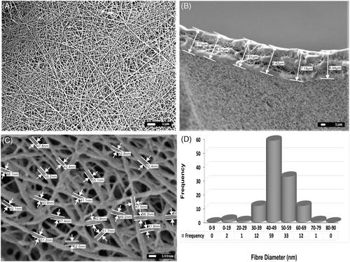
Cell replacement therapy is under development for dry age-related macular degeneration (AMD). A thin membrane resembling the Bruch's membrane is required to form a cell-on-membrane construct with retinal pigment epithelial (RPE) cells. These cells have been differentiated from human embryonic stem cells (hESCs) in vitro. A carrier membrane is required for cell implantation, which is biocompatible for cell growth and has dimensions and physical properties resembling the Bruch's membrane. Here a nanofiber electrospun poly-L-lactic acid (PLLA) membrane is tested for capacity to support cell growth and maturation. The requirements for laminin coating of the membrane are identified here. A porous electrospun nanofibrous PLLA membrane of ∼50 nm fiber diameter was developed as a prototype support for functional RPE cells grown as a monolayer. The need for laminin coating applied to the membrane following treatment with poly-L-ornithine (PLO), was identified in terms of cell growth and survival. Test membranes were compared in terms of hydrophilicity after laminin coating, mechanical properties of surface roughness and Young's modulus, porosity and ability to promote the attachment and proliferation of hESC-RPE cells in culture for up to 8 weeks. Over this time, RPE cell proliferation, morphology, and marker and gene expression, were monitored. The functional capacity of cell monolayers was identified in terms of transepithelial electrical resistance (TEER), phagocytosis of cells, as well as expression of the cytokines, vascular endothelial growth factor (VEGF) and pigment epithelium-derived factor (PEDF). PLLA polymer fibers are naturally hydrophobic, so their hydrophilicity was improved by pretreatment with PLO for subsequent coating with the bioactive protein laminin. They were then assessed for amount of laminin adsorbed, contact angle and uniformity of coating using scanning electron microscopy (SEM). Pretreatment with 100% PLO gave the best result over 10% PLO treatment or no treatment prior to laminin adsorption with significantly greater surface stiffness and modulus. By 6 weeks after cell plating, the coated membranes could support a mature RPE monolayer showing a dense apical microvillus structure and pigmented 3D polygonal cell morphology. After 8 weeks, PLO (100%)-Lam coated membranes exhibited the highest cell number, cell proliferation, and RPE barrier function measured as TEER. RPE cells showed the higher levels of specific surface marker and gene expression. Microphthalmia-associated transcription factor expression was highly upregulated indicating maturation of cells. Functionality of cells was indicated by expression of VEGF and PEDF genes as well as phagocytic capacity. In conclusion, electrospun PLLA membranes coated with PLO-Lam have the physical and biological properties to support the distribution and migration of hESC-RPE cells throughout the whole structure. They represent a good membrane candidate for preparation of hESC-RPE cells as a monolayer for implantation into the subretinal space of AMD patients.
期刊介绍:
The Journal of Biomedical Materials Research Part A is an international, interdisciplinary, English-language publication of original contributions concerning studies of the preparation, performance, and evaluation of biomaterials; the chemical, physical, toxicological, and mechanical behavior of materials in physiological environments; and the response of blood and tissues to biomaterials. The Journal publishes peer-reviewed articles on all relevant biomaterial topics including the science and technology of alloys,polymers, ceramics, and reprocessed animal and human tissues in surgery,dentistry, artificial organs, and other medical devices. The Journal also publishes articles in interdisciplinary areas such as tissue engineering and controlled release technology where biomaterials play a significant role in the performance of the medical device.
The Journal of Biomedical Materials Research is the official journal of the Society for Biomaterials (USA), the Japanese Society for Biomaterials, the Australasian Society for Biomaterials, and the Korean Society for Biomaterials.
Articles are welcomed from all scientists. Membership in the Society for Biomaterials is not a prerequisite for submission.


 分享
分享
 求助内容:
求助内容: 应助结果提醒方式:
 扫码关注我们
