Asmita Karki, Bishan Man Thaiba, K. C. Shishir Acharya, Thakur Sedai, Baburam Kandel, Hari Paudyal, Khaga Raj Sharma, Basant Giri, Bhanu Bhakta Neupane
{"title":"智能手机显微镜成像和量化饮用水中微塑料的方法。","authors":"Asmita Karki, Bishan Man Thaiba, K. C. Shishir Acharya, Thakur Sedai, Baburam Kandel, Hari Paudyal, Khaga Raj Sharma, Basant Giri, Bhanu Bhakta Neupane","doi":"10.1002/jemt.24596","DOIUrl":null,"url":null,"abstract":"<div>\n \n \n <section>\n \n <p>Analysis of microplastics in drinking water is often challenging due to smaller particle size and low particle count. In this study, we used a low cost and an easy to assemble smartphone microscopic system for imaging and quantitating microplastic particles as small as 20 μm. The system consisted of a spherical sapphire ball lens of 4 mm diameter attached to a smartphone camera as a major imaging component. It also involved pre-concentration of the sample using ZnCl<sub>2</sub> solution. The spike recovery and limit of detection of the method in filtered distilled and deionized water samples (<i>n</i> = 9) were 55.6% ± 9.7% and 34 particles/L, respectively. Imaging performance of the microscopic system was similar to a commercial bright field microscopic system. The method was further implemented to examine microplastic particles in commercial bottled and jar water samples (<i>n</i> = 20). The particles count in bottled and jar water samples ranged from 0–91 particles/L to 0–130 particles/L, respectively. In both sample types, particles of diverse shape and size were observed. The particles collected from water samples were further confirmed by FTIR spectra (<i>n</i> = 36), which found 97% of the particles tested were made of plastic material. These findings suggested that the smartphone microscopic system can be implemented as a low-cost alternative for preliminary screening of microplastic in drinking water samples.</p>\n </section>\n \n <section>\n \n <h3> Research Highlights</h3>\n \n <div>\n <ul>\n \n <li>Ball lens based smartphone microscopic method was used for microplastic analysis.</li>\n \n <li>Particles of diverse shape and size were found in bottle and jar water samples.</li>\n </ul>\n </div>\n </section>\n </div>","PeriodicalId":18684,"journal":{"name":"Microscopy Research and Technique","volume":null,"pages":null},"PeriodicalIF":2.0000,"publicationDate":"2024-05-11","publicationTypes":"Journal Article","fieldsOfStudy":null,"isOpenAccess":false,"openAccessPdf":"","citationCount":"0","resultStr":"{\"title\":\"Smartphone microscopic method for imaging and quantification of microplastics in drinking water\",\"authors\":\"Asmita Karki, Bishan Man Thaiba, K. C. Shishir Acharya, Thakur Sedai, Baburam Kandel, Hari Paudyal, Khaga Raj Sharma, Basant Giri, Bhanu Bhakta Neupane\",\"doi\":\"10.1002/jemt.24596\",\"DOIUrl\":null,\"url\":null,\"abstract\":\"<div>\\n \\n \\n <section>\\n \\n <p>Analysis of microplastics in drinking water is often challenging due to smaller particle size and low particle count. In this study, we used a low cost and an easy to assemble smartphone microscopic system for imaging and quantitating microplastic particles as small as 20 μm. The system consisted of a spherical sapphire ball lens of 4 mm diameter attached to a smartphone camera as a major imaging component. It also involved pre-concentration of the sample using ZnCl<sub>2</sub> solution. The spike recovery and limit of detection of the method in filtered distilled and deionized water samples (<i>n</i> = 9) were 55.6% ± 9.7% and 34 particles/L, respectively. Imaging performance of the microscopic system was similar to a commercial bright field microscopic system. The method was further implemented to examine microplastic particles in commercial bottled and jar water samples (<i>n</i> = 20). The particles count in bottled and jar water samples ranged from 0–91 particles/L to 0–130 particles/L, respectively. In both sample types, particles of diverse shape and size were observed. The particles collected from water samples were further confirmed by FTIR spectra (<i>n</i> = 36), which found 97% of the particles tested were made of plastic material. These findings suggested that the smartphone microscopic system can be implemented as a low-cost alternative for preliminary screening of microplastic in drinking water samples.</p>\\n </section>\\n \\n <section>\\n \\n <h3> Research Highlights</h3>\\n \\n <div>\\n <ul>\\n \\n <li>Ball lens based smartphone microscopic method was used for microplastic analysis.</li>\\n \\n <li>Particles of diverse shape and size were found in bottle and jar water samples.</li>\\n </ul>\\n </div>\\n </section>\\n </div>\",\"PeriodicalId\":18684,\"journal\":{\"name\":\"Microscopy Research and Technique\",\"volume\":null,\"pages\":null},\"PeriodicalIF\":2.0000,\"publicationDate\":\"2024-05-11\",\"publicationTypes\":\"Journal Article\",\"fieldsOfStudy\":null,\"isOpenAccess\":false,\"openAccessPdf\":\"\",\"citationCount\":\"0\",\"resultStr\":null,\"platform\":\"Semanticscholar\",\"paperid\":null,\"PeriodicalName\":\"Microscopy Research and Technique\",\"FirstCategoryId\":\"5\",\"ListUrlMain\":\"https://onlinelibrary.wiley.com/doi/10.1002/jemt.24596\",\"RegionNum\":3,\"RegionCategory\":\"工程技术\",\"ArticlePicture\":[],\"TitleCN\":null,\"AbstractTextCN\":null,\"PMCID\":null,\"EPubDate\":\"\",\"PubModel\":\"\",\"JCR\":\"Q2\",\"JCRName\":\"ANATOMY & MORPHOLOGY\",\"Score\":null,\"Total\":0}","platform":"Semanticscholar","paperid":null,"PeriodicalName":"Microscopy Research and Technique","FirstCategoryId":"5","ListUrlMain":"https://onlinelibrary.wiley.com/doi/10.1002/jemt.24596","RegionNum":3,"RegionCategory":"工程技术","ArticlePicture":[],"TitleCN":null,"AbstractTextCN":null,"PMCID":null,"EPubDate":"","PubModel":"","JCR":"Q2","JCRName":"ANATOMY & MORPHOLOGY","Score":null,"Total":0}
Smartphone microscopic method for imaging and quantification of microplastics in drinking water
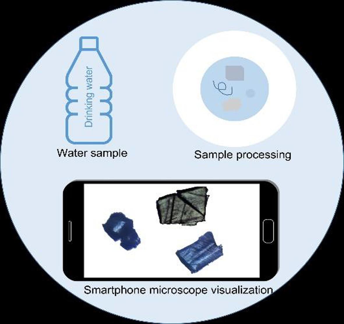
Analysis of microplastics in drinking water is often challenging due to smaller particle size and low particle count. In this study, we used a low cost and an easy to assemble smartphone microscopic system for imaging and quantitating microplastic particles as small as 20 μm. The system consisted of a spherical sapphire ball lens of 4 mm diameter attached to a smartphone camera as a major imaging component. It also involved pre-concentration of the sample using ZnCl2 solution. The spike recovery and limit of detection of the method in filtered distilled and deionized water samples (n = 9) were 55.6% ± 9.7% and 34 particles/L, respectively. Imaging performance of the microscopic system was similar to a commercial bright field microscopic system. The method was further implemented to examine microplastic particles in commercial bottled and jar water samples (n = 20). The particles count in bottled and jar water samples ranged from 0–91 particles/L to 0–130 particles/L, respectively. In both sample types, particles of diverse shape and size were observed. The particles collected from water samples were further confirmed by FTIR spectra (n = 36), which found 97% of the particles tested were made of plastic material. These findings suggested that the smartphone microscopic system can be implemented as a low-cost alternative for preliminary screening of microplastic in drinking water samples.
Research Highlights
Ball lens based smartphone microscopic method was used for microplastic analysis.
Particles of diverse shape and size were found in bottle and jar water samples.
期刊介绍:
Microscopy Research and Technique (MRT) publishes articles on all aspects of advanced microscopy original architecture and methodologies with applications in the biological, clinical, chemical, and materials sciences. Original basic and applied research as well as technical papers dealing with the various subsets of microscopy are encouraged. MRT is the right form for those developing new microscopy methods or using the microscope to answer key questions in basic and applied research.


 分享
分享
 求助内容:
求助内容: 应助结果提醒方式:
 扫码关注我们
