{"title":"利用智能成像流式细胞仪诊断浆液性积液","authors":"Mengping Long, Yueyun Weng, Liye Mei, Dingchao Yang, Shubin Wei, Guanxiong Meng, Wanyue Zhao, Sheng Liu, Du Wang, Yiqiang Liu, Hui Shen, Jianxuan Hou, Yu Xu, Liang Tao, Fuling Zhou, Hongwei Chen, Taobo Hu, Cheng Lei","doi":"10.1002/adsr.202300183","DOIUrl":null,"url":null,"abstract":"<p>A serous effusion is a buildup of extra fluid in the serous cavities including pleural, peritoneal, and pericardial cavities. It is important to distinguish benign reactive effusions from effusions caused by malignant proliferation in cytopathology since different diagnoses can lead to completely different disease staging and therapeutic choices. The conventional cytopathology procedure has the disadvantages of low throughput and low objectivity. To enhance the efficiency and accuracy of malignant serous effusion diagnosis, in this paper, an imaging flow cytometry, called optofluidic time-stretch microscopy is first employed, to image the cells in the serous effusion at an event rate of 100 000 events per second and with a spatial resolution better than 1 µm. The acquired cellular images are then analyzed using a convolutional neural network, by which the malignant cells are accurately detected. The performance of the method is validated with 18 clinical samples, including 14 malignant and 4 benign ones. The results show that the method can detect malignant cells at an accuracy of 90.53%. The high throughput, high accuracy, and high convenience of the method make it a potential solution for malignant serous effusion diagnosis in various scenarios.</p>","PeriodicalId":100037,"journal":{"name":"Advanced Sensor Research","volume":"3 8","pages":""},"PeriodicalIF":5.0000,"publicationDate":"2024-05-13","publicationTypes":"Journal Article","fieldsOfStudy":null,"isOpenAccess":false,"openAccessPdf":"https://onlinelibrary.wiley.com/doi/epdf/10.1002/adsr.202300183","citationCount":"0","resultStr":"{\"title\":\"Diagnosis of Serous Effusion with Intelligent Imaging Flow Cytometry\",\"authors\":\"Mengping Long, Yueyun Weng, Liye Mei, Dingchao Yang, Shubin Wei, Guanxiong Meng, Wanyue Zhao, Sheng Liu, Du Wang, Yiqiang Liu, Hui Shen, Jianxuan Hou, Yu Xu, Liang Tao, Fuling Zhou, Hongwei Chen, Taobo Hu, Cheng Lei\",\"doi\":\"10.1002/adsr.202300183\",\"DOIUrl\":null,\"url\":null,\"abstract\":\"<p>A serous effusion is a buildup of extra fluid in the serous cavities including pleural, peritoneal, and pericardial cavities. It is important to distinguish benign reactive effusions from effusions caused by malignant proliferation in cytopathology since different diagnoses can lead to completely different disease staging and therapeutic choices. The conventional cytopathology procedure has the disadvantages of low throughput and low objectivity. To enhance the efficiency and accuracy of malignant serous effusion diagnosis, in this paper, an imaging flow cytometry, called optofluidic time-stretch microscopy is first employed, to image the cells in the serous effusion at an event rate of 100 000 events per second and with a spatial resolution better than 1 µm. The acquired cellular images are then analyzed using a convolutional neural network, by which the malignant cells are accurately detected. The performance of the method is validated with 18 clinical samples, including 14 malignant and 4 benign ones. The results show that the method can detect malignant cells at an accuracy of 90.53%. The high throughput, high accuracy, and high convenience of the method make it a potential solution for malignant serous effusion diagnosis in various scenarios.</p>\",\"PeriodicalId\":100037,\"journal\":{\"name\":\"Advanced Sensor Research\",\"volume\":\"3 8\",\"pages\":\"\"},\"PeriodicalIF\":5.0000,\"publicationDate\":\"2024-05-13\",\"publicationTypes\":\"Journal Article\",\"fieldsOfStudy\":null,\"isOpenAccess\":false,\"openAccessPdf\":\"https://onlinelibrary.wiley.com/doi/epdf/10.1002/adsr.202300183\",\"citationCount\":\"0\",\"resultStr\":null,\"platform\":\"Semanticscholar\",\"paperid\":null,\"PeriodicalName\":\"Advanced Sensor Research\",\"FirstCategoryId\":\"1085\",\"ListUrlMain\":\"https://advanced.onlinelibrary.wiley.com/doi/10.1002/adsr.202300183\",\"RegionNum\":0,\"RegionCategory\":null,\"ArticlePicture\":[],\"TitleCN\":null,\"AbstractTextCN\":null,\"PMCID\":null,\"EPubDate\":\"\",\"PubModel\":\"\",\"JCR\":\"\",\"JCRName\":\"\",\"Score\":null,\"Total\":0}","platform":"Semanticscholar","paperid":null,"PeriodicalName":"Advanced Sensor Research","FirstCategoryId":"1085","ListUrlMain":"https://advanced.onlinelibrary.wiley.com/doi/10.1002/adsr.202300183","RegionNum":0,"RegionCategory":null,"ArticlePicture":[],"TitleCN":null,"AbstractTextCN":null,"PMCID":null,"EPubDate":"","PubModel":"","JCR":"","JCRName":"","Score":null,"Total":0}
Diagnosis of Serous Effusion with Intelligent Imaging Flow Cytometry
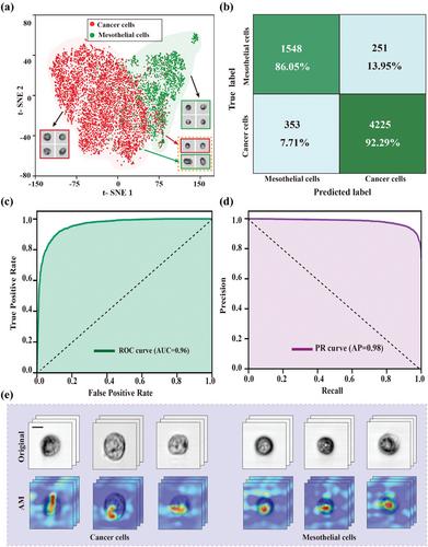
A serous effusion is a buildup of extra fluid in the serous cavities including pleural, peritoneal, and pericardial cavities. It is important to distinguish benign reactive effusions from effusions caused by malignant proliferation in cytopathology since different diagnoses can lead to completely different disease staging and therapeutic choices. The conventional cytopathology procedure has the disadvantages of low throughput and low objectivity. To enhance the efficiency and accuracy of malignant serous effusion diagnosis, in this paper, an imaging flow cytometry, called optofluidic time-stretch microscopy is first employed, to image the cells in the serous effusion at an event rate of 100 000 events per second and with a spatial resolution better than 1 µm. The acquired cellular images are then analyzed using a convolutional neural network, by which the malignant cells are accurately detected. The performance of the method is validated with 18 clinical samples, including 14 malignant and 4 benign ones. The results show that the method can detect malignant cells at an accuracy of 90.53%. The high throughput, high accuracy, and high convenience of the method make it a potential solution for malignant serous effusion diagnosis in various scenarios.


 分享
分享

 求助内容:
求助内容: 应助结果提醒方式:
 扫码关注我们
