{"title":"FAPI 和 FDG PET/CT 定量参数在非小细胞肺癌纵隔淋巴结转移检测中的诊断价值比较","authors":"Min Wang, Jiayu Zhang, Bin Wu, Chunyin Zhang","doi":"10.1007/s40336-024-00644-1","DOIUrl":null,"url":null,"abstract":"<h3 data-test=\"abstract-sub-heading\">Objective</h3><p>This study explores the diagnostic value of quantitative parameters from <sup>68</sup>Ga-FAPI-04 PET/CT and <sup>18</sup>F-FDG PET/CT in the detection of mediastinal lymph node metastasis in non-small cell lung cancer (NSCLC) patients.</p><h3 data-test=\"abstract-sub-heading\">Methods</h3><p>Seventeen NSCLC patients patients undergoing imaging with FAPI and FDG were included in the study. Measurements were taken for short diameter, long diameter, density of mediastinal lymph nodes, as well as SUVmax of mediastinal blood pool (MBP- SUVmax), primary tumor (PT-SUVmax), and lymph nodes (LN-SUVmax) in both imaging modalities. LN-SUVmax / MBP- SUVmax, LN-SUVmax / PT-SUVmax, and LN-SUVmax /short diameter ratios were calculated for lymph nodes. Statistical differences between the parameters of imaging modalities in the mediastinal lymph node metastasis group and the non-metastasis group were analyzed. Statistical differences between FAPI and FDG imaging parameters were also compared. Receiver Operating Characteristic (ROC) curves were utilized to determine optimal diagnostic thresholds, along with corresponding sensitivity, specificity, and area under the curve (AUC) for each parameter in the detection of mediastinal lymph node metastasis.</p><h3 data-test=\"abstract-sub-heading\">Results</h3><p>In FAPI imaging, LN-SUVmax, LN-SUVmax / MBP- SUVmax, LN-SUVmax / PT-SUVmax, and LN-SUVmax /short diameter were higher in the mediastinal lymph node metastasis group compared to the non-metastasis group, with statistical significance (<i>p</i> < 0.05). In FDG imaging, only LN-SUVmax / PT-SUVmax was higher in the mediastinal lymph node metastasis group, with statistical significance (<i>p</i> < 0.05). Two parameters (LN-SUVmax/MBP-SUVmax, LN-SUVmax/PT-SUVmax) in the FAPI group and three parameters (LN-SUVmax, LN-SUVmax/MBP-SUVmax, LN-SUVmax/short diameter) in the FDG group showed positive and negative correlations, respectively, in the mediastinal lymph node metastasis and non-metastasis groups, all with statistical significance (<i>p</i> < 0.05). The AUC of FAPI imaging for LN-SUVmax (0.907 vs. 0.667) and LN-SUVmax/PT- SUVmax (0.772 vs. 0.704) were higher compared to FDG imaging, with statistical significance (<i>p</i> < 0.05).</p><h3 data-test=\"abstract-sub-heading\">Conclusion</h3><p><sup>68</sup>Ga-FAPI-04 PET/CT imaging exhibits a certain advantage over <sup>18</sup>F-FDG PET/CT in the detection of mediastinal lymph node metastasis in NSCLC patients, suggesting potential clinical value in the staging of NSCLC patients.</p>","PeriodicalId":48600,"journal":{"name":"Clinical and Translational Imaging","volume":"10 1","pages":""},"PeriodicalIF":1.6000,"publicationDate":"2024-05-27","publicationTypes":"Journal Article","fieldsOfStudy":null,"isOpenAccess":false,"openAccessPdf":"","citationCount":"0","resultStr":"{\"title\":\"Comparison of diagnostic value of quantitative parameters from FAPI and FDG PET/CT in the detection of mediastinal lymph node metastasis in non-small cell lung cancer\",\"authors\":\"Min Wang, Jiayu Zhang, Bin Wu, Chunyin Zhang\",\"doi\":\"10.1007/s40336-024-00644-1\",\"DOIUrl\":null,\"url\":null,\"abstract\":\"<h3 data-test=\\\"abstract-sub-heading\\\">Objective</h3><p>This study explores the diagnostic value of quantitative parameters from <sup>68</sup>Ga-FAPI-04 PET/CT and <sup>18</sup>F-FDG PET/CT in the detection of mediastinal lymph node metastasis in non-small cell lung cancer (NSCLC) patients.</p><h3 data-test=\\\"abstract-sub-heading\\\">Methods</h3><p>Seventeen NSCLC patients patients undergoing imaging with FAPI and FDG were included in the study. Measurements were taken for short diameter, long diameter, density of mediastinal lymph nodes, as well as SUVmax of mediastinal blood pool (MBP- SUVmax), primary tumor (PT-SUVmax), and lymph nodes (LN-SUVmax) in both imaging modalities. LN-SUVmax / MBP- SUVmax, LN-SUVmax / PT-SUVmax, and LN-SUVmax /short diameter ratios were calculated for lymph nodes. Statistical differences between the parameters of imaging modalities in the mediastinal lymph node metastasis group and the non-metastasis group were analyzed. Statistical differences between FAPI and FDG imaging parameters were also compared. Receiver Operating Characteristic (ROC) curves were utilized to determine optimal diagnostic thresholds, along with corresponding sensitivity, specificity, and area under the curve (AUC) for each parameter in the detection of mediastinal lymph node metastasis.</p><h3 data-test=\\\"abstract-sub-heading\\\">Results</h3><p>In FAPI imaging, LN-SUVmax, LN-SUVmax / MBP- SUVmax, LN-SUVmax / PT-SUVmax, and LN-SUVmax /short diameter were higher in the mediastinal lymph node metastasis group compared to the non-metastasis group, with statistical significance (<i>p</i> < 0.05). In FDG imaging, only LN-SUVmax / PT-SUVmax was higher in the mediastinal lymph node metastasis group, with statistical significance (<i>p</i> < 0.05). Two parameters (LN-SUVmax/MBP-SUVmax, LN-SUVmax/PT-SUVmax) in the FAPI group and three parameters (LN-SUVmax, LN-SUVmax/MBP-SUVmax, LN-SUVmax/short diameter) in the FDG group showed positive and negative correlations, respectively, in the mediastinal lymph node metastasis and non-metastasis groups, all with statistical significance (<i>p</i> < 0.05). The AUC of FAPI imaging for LN-SUVmax (0.907 vs. 0.667) and LN-SUVmax/PT- SUVmax (0.772 vs. 0.704) were higher compared to FDG imaging, with statistical significance (<i>p</i> < 0.05).</p><h3 data-test=\\\"abstract-sub-heading\\\">Conclusion</h3><p><sup>68</sup>Ga-FAPI-04 PET/CT imaging exhibits a certain advantage over <sup>18</sup>F-FDG PET/CT in the detection of mediastinal lymph node metastasis in NSCLC patients, suggesting potential clinical value in the staging of NSCLC patients.</p>\",\"PeriodicalId\":48600,\"journal\":{\"name\":\"Clinical and Translational Imaging\",\"volume\":\"10 1\",\"pages\":\"\"},\"PeriodicalIF\":1.6000,\"publicationDate\":\"2024-05-27\",\"publicationTypes\":\"Journal Article\",\"fieldsOfStudy\":null,\"isOpenAccess\":false,\"openAccessPdf\":\"\",\"citationCount\":\"0\",\"resultStr\":null,\"platform\":\"Semanticscholar\",\"paperid\":null,\"PeriodicalName\":\"Clinical and Translational Imaging\",\"FirstCategoryId\":\"3\",\"ListUrlMain\":\"https://doi.org/10.1007/s40336-024-00644-1\",\"RegionNum\":4,\"RegionCategory\":\"医学\",\"ArticlePicture\":[],\"TitleCN\":null,\"AbstractTextCN\":null,\"PMCID\":null,\"EPubDate\":\"\",\"PubModel\":\"\",\"JCR\":\"Q2\",\"JCRName\":\"RADIOLOGY, NUCLEAR MEDICINE & MEDICAL IMAGING\",\"Score\":null,\"Total\":0}","platform":"Semanticscholar","paperid":null,"PeriodicalName":"Clinical and Translational Imaging","FirstCategoryId":"3","ListUrlMain":"https://doi.org/10.1007/s40336-024-00644-1","RegionNum":4,"RegionCategory":"医学","ArticlePicture":[],"TitleCN":null,"AbstractTextCN":null,"PMCID":null,"EPubDate":"","PubModel":"","JCR":"Q2","JCRName":"RADIOLOGY, NUCLEAR MEDICINE & MEDICAL IMAGING","Score":null,"Total":0}
Comparison of diagnostic value of quantitative parameters from FAPI and FDG PET/CT in the detection of mediastinal lymph node metastasis in non-small cell lung cancer
Objective
This study explores the diagnostic value of quantitative parameters from 68Ga-FAPI-04 PET/CT and 18F-FDG PET/CT in the detection of mediastinal lymph node metastasis in non-small cell lung cancer (NSCLC) patients.
Methods
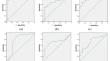
Seventeen NSCLC patients patients undergoing imaging with FAPI and FDG were included in the study. Measurements were taken for short diameter, long diameter, density of mediastinal lymph nodes, as well as SUVmax of mediastinal blood pool (MBP- SUVmax), primary tumor (PT-SUVmax), and lymph nodes (LN-SUVmax) in both imaging modalities. LN-SUVmax / MBP- SUVmax, LN-SUVmax / PT-SUVmax, and LN-SUVmax /short diameter ratios were calculated for lymph nodes. Statistical differences between the parameters of imaging modalities in the mediastinal lymph node metastasis group and the non-metastasis group were analyzed. Statistical differences between FAPI and FDG imaging parameters were also compared. Receiver Operating Characteristic (ROC) curves were utilized to determine optimal diagnostic thresholds, along with corresponding sensitivity, specificity, and area under the curve (AUC) for each parameter in the detection of mediastinal lymph node metastasis.
Results
In FAPI imaging, LN-SUVmax, LN-SUVmax / MBP- SUVmax, LN-SUVmax / PT-SUVmax, and LN-SUVmax /short diameter were higher in the mediastinal lymph node metastasis group compared to the non-metastasis group, with statistical significance (p < 0.05). In FDG imaging, only LN-SUVmax / PT-SUVmax was higher in the mediastinal lymph node metastasis group, with statistical significance (p < 0.05). Two parameters (LN-SUVmax/MBP-SUVmax, LN-SUVmax/PT-SUVmax) in the FAPI group and three parameters (LN-SUVmax, LN-SUVmax/MBP-SUVmax, LN-SUVmax/short diameter) in the FDG group showed positive and negative correlations, respectively, in the mediastinal lymph node metastasis and non-metastasis groups, all with statistical significance (p < 0.05). The AUC of FAPI imaging for LN-SUVmax (0.907 vs. 0.667) and LN-SUVmax/PT- SUVmax (0.772 vs. 0.704) were higher compared to FDG imaging, with statistical significance (p < 0.05).
Conclusion
68Ga-FAPI-04 PET/CT imaging exhibits a certain advantage over 18F-FDG PET/CT in the detection of mediastinal lymph node metastasis in NSCLC patients, suggesting potential clinical value in the staging of NSCLC patients.
期刊介绍:
Clinical and Translational Imaging is an international journal that publishes timely, up-to-date summaries on clinical practice and translational research and clinical applications of approved and experimental radiopharmaceuticals for diagnostic and therapeutic purposes. Coverage includes such topics as advanced preclinical evidence in the fields of physics, dosimetry, radiation biology and radiopharmacy with relevance to applications in human subjects. The journal benefits a readership of nuclear medicine practitioners and allied professionals involved in molecular imaging and therapy.


 分享
分享
 求助内容:
求助内容: 应助结果提醒方式:
 扫码关注我们
