{"title":"预切乳头切开术后胆道口的可见度:白光成像与纹理和色彩增强成像的比较。","authors":"Haruka Toyonaga, Toshifumi Kin, Hajime Yamazaki, Ryo Ando, Kosuke Iwano, Risa Nakamura, Tatsuya Ishii, Tsuyoshi Hayashi, Kuniyuki Takahashi, Akio Katanuma","doi":"10.1002/jhbp.12005","DOIUrl":null,"url":null,"abstract":"<div>\n \n \n <section>\n \n <h3> Background</h3>\n \n <p>Precut papillotomy is performed in cases of difficult biliary cannulation, but identification of the biliary orifice is difficult. Texture and color enhancement imaging (TXI) can enhance the structure, color, and brightness. This study compared TXI and white light imaging (WLI) in visibility of biliary orifices.</p>\n </section>\n \n <section>\n \n <h3> Methods</h3>\n \n <p>We retrospectively examined 20 patients who underwent bile duct cannulation using both WLI and TXI after precut papillotomy at our center between 2021 and 2022. On WLI and TXI images displayed in random order, bile duct orifice on precut-incision surface of each image was independently evaluated by eight evaluators. Single-indication accuracy rate of biliary orifices, visibility score rated on a 4-grade scale, and color difference between the biliary orifice and the surrounding tissue were examined.</p>\n </section>\n \n <section>\n \n <h3> Results</h3>\n \n <p>The single-indication accuracy rate was higher in TXI compared to WLI (50.6% vs. 35.6%, odds ratio 2.26 [95% CI: 1.32–3.89], <i>p</i> = .003). The time to indicate the biliary orifice was comparable between TXI and WLI (median, 9.7 s [range, 2.6–43] vs. 10.9 s [1.5–64], <i>p</i> = .086). Furthermore, the visibility score was higher in TXI than in WLI (median, 3 [interquartile range, 2–3] vs. 2 [2, 3], <i>p</i> < .001), and the color difference between the biliary orifice and surrounding tissue in TXI was more pronounced than in WLI (median, 22.9 [range, 9.39–55.2] vs. 18.0 [6.48–43.0]; <i>p</i> < .001).</p>\n </section>\n \n <section>\n \n <h3> Conclusions</h3>\n \n <p>TXI enhanced the color difference and visibility of the biliary orifice after precut and improved single-indication accuracy rate, suggesting that it could be useful for biliary cannulation after precut papillotomy.</p>\n </section>\n </div>","PeriodicalId":16056,"journal":{"name":"Journal of Hepato‐Biliary‐Pancreatic Sciences","volume":"31 8","pages":"591-600"},"PeriodicalIF":2.5000,"publicationDate":"2024-06-02","publicationTypes":"Journal Article","fieldsOfStudy":null,"isOpenAccess":false,"openAccessPdf":"","citationCount":"0","resultStr":"{\"title\":\"Visibility of the biliary orifice after precut papillotomy: Comparison between white light imaging and texture and color enhancement imaging\",\"authors\":\"Haruka Toyonaga, Toshifumi Kin, Hajime Yamazaki, Ryo Ando, Kosuke Iwano, Risa Nakamura, Tatsuya Ishii, Tsuyoshi Hayashi, Kuniyuki Takahashi, Akio Katanuma\",\"doi\":\"10.1002/jhbp.12005\",\"DOIUrl\":null,\"url\":null,\"abstract\":\"<div>\\n \\n \\n <section>\\n \\n <h3> Background</h3>\\n \\n <p>Precut papillotomy is performed in cases of difficult biliary cannulation, but identification of the biliary orifice is difficult. Texture and color enhancement imaging (TXI) can enhance the structure, color, and brightness. This study compared TXI and white light imaging (WLI) in visibility of biliary orifices.</p>\\n </section>\\n \\n <section>\\n \\n <h3> Methods</h3>\\n \\n <p>We retrospectively examined 20 patients who underwent bile duct cannulation using both WLI and TXI after precut papillotomy at our center between 2021 and 2022. On WLI and TXI images displayed in random order, bile duct orifice on precut-incision surface of each image was independently evaluated by eight evaluators. Single-indication accuracy rate of biliary orifices, visibility score rated on a 4-grade scale, and color difference between the biliary orifice and the surrounding tissue were examined.</p>\\n </section>\\n \\n <section>\\n \\n <h3> Results</h3>\\n \\n <p>The single-indication accuracy rate was higher in TXI compared to WLI (50.6% vs. 35.6%, odds ratio 2.26 [95% CI: 1.32–3.89], <i>p</i> = .003). The time to indicate the biliary orifice was comparable between TXI and WLI (median, 9.7 s [range, 2.6–43] vs. 10.9 s [1.5–64], <i>p</i> = .086). Furthermore, the visibility score was higher in TXI than in WLI (median, 3 [interquartile range, 2–3] vs. 2 [2, 3], <i>p</i> < .001), and the color difference between the biliary orifice and surrounding tissue in TXI was more pronounced than in WLI (median, 22.9 [range, 9.39–55.2] vs. 18.0 [6.48–43.0]; <i>p</i> < .001).</p>\\n </section>\\n \\n <section>\\n \\n <h3> Conclusions</h3>\\n \\n <p>TXI enhanced the color difference and visibility of the biliary orifice after precut and improved single-indication accuracy rate, suggesting that it could be useful for biliary cannulation after precut papillotomy.</p>\\n </section>\\n </div>\",\"PeriodicalId\":16056,\"journal\":{\"name\":\"Journal of Hepato‐Biliary‐Pancreatic Sciences\",\"volume\":\"31 8\",\"pages\":\"591-600\"},\"PeriodicalIF\":2.5000,\"publicationDate\":\"2024-06-02\",\"publicationTypes\":\"Journal Article\",\"fieldsOfStudy\":null,\"isOpenAccess\":false,\"openAccessPdf\":\"\",\"citationCount\":\"0\",\"resultStr\":null,\"platform\":\"Semanticscholar\",\"paperid\":null,\"PeriodicalName\":\"Journal of Hepato‐Biliary‐Pancreatic Sciences\",\"FirstCategoryId\":\"3\",\"ListUrlMain\":\"https://onlinelibrary.wiley.com/doi/10.1002/jhbp.12005\",\"RegionNum\":3,\"RegionCategory\":\"医学\",\"ArticlePicture\":[],\"TitleCN\":null,\"AbstractTextCN\":null,\"PMCID\":null,\"EPubDate\":\"\",\"PubModel\":\"\",\"JCR\":\"Q2\",\"JCRName\":\"GASTROENTEROLOGY & HEPATOLOGY\",\"Score\":null,\"Total\":0}","platform":"Semanticscholar","paperid":null,"PeriodicalName":"Journal of Hepato‐Biliary‐Pancreatic Sciences","FirstCategoryId":"3","ListUrlMain":"https://onlinelibrary.wiley.com/doi/10.1002/jhbp.12005","RegionNum":3,"RegionCategory":"医学","ArticlePicture":[],"TitleCN":null,"AbstractTextCN":null,"PMCID":null,"EPubDate":"","PubModel":"","JCR":"Q2","JCRName":"GASTROENTEROLOGY & HEPATOLOGY","Score":null,"Total":0}
Visibility of the biliary orifice after precut papillotomy: Comparison between white light imaging and texture and color enhancement imaging
Background
Precut papillotomy is performed in cases of difficult biliary cannulation, but identification of the biliary orifice is difficult. Texture and color enhancement imaging (TXI) can enhance the structure, color, and brightness. This study compared TXI and white light imaging (WLI) in visibility of biliary orifices.
Methods
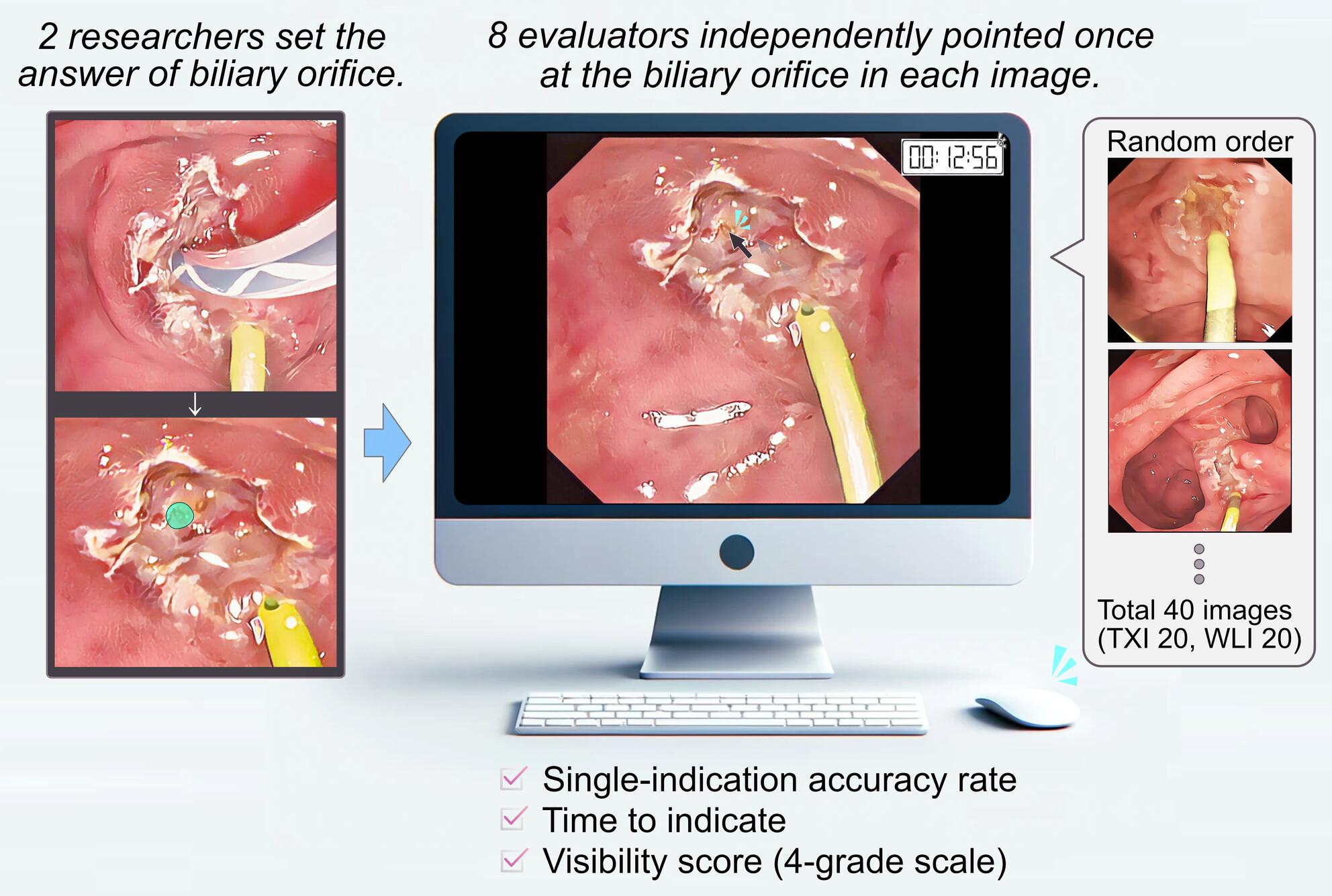
We retrospectively examined 20 patients who underwent bile duct cannulation using both WLI and TXI after precut papillotomy at our center between 2021 and 2022. On WLI and TXI images displayed in random order, bile duct orifice on precut-incision surface of each image was independently evaluated by eight evaluators. Single-indication accuracy rate of biliary orifices, visibility score rated on a 4-grade scale, and color difference between the biliary orifice and the surrounding tissue were examined.
Results
The single-indication accuracy rate was higher in TXI compared to WLI (50.6% vs. 35.6%, odds ratio 2.26 [95% CI: 1.32–3.89], p = .003). The time to indicate the biliary orifice was comparable between TXI and WLI (median, 9.7 s [range, 2.6–43] vs. 10.9 s [1.5–64], p = .086). Furthermore, the visibility score was higher in TXI than in WLI (median, 3 [interquartile range, 2–3] vs. 2 [2, 3], p < .001), and the color difference between the biliary orifice and surrounding tissue in TXI was more pronounced than in WLI (median, 22.9 [range, 9.39–55.2] vs. 18.0 [6.48–43.0]; p < .001).
Conclusions
TXI enhanced the color difference and visibility of the biliary orifice after precut and improved single-indication accuracy rate, suggesting that it could be useful for biliary cannulation after precut papillotomy.
期刊介绍:
The Journal of Hepato-Biliary-Pancreatic Sciences (JHBPS) is the leading peer-reviewed journal in the field of hepato-biliary-pancreatic sciences. JHBPS publishes articles dealing with clinical research as well as translational research on all aspects of this field. Coverage includes Original Article, Review Article, Images of Interest, Rapid Communication and an announcement section. Letters to the Editor and comments on the journal’s policies or content are also included. JHBPS welcomes submissions from surgeons, physicians, endoscopists, radiologists, oncologists, and pathologists.


 分享
分享
 求助内容:
求助内容: 应助结果提醒方式:
 扫码关注我们
