{"title":"脑室下肿瘤儿科患者的皮层和皮层下结构变化。","authors":"Barış Genç, Kerim Aslan, Derya Bako, Semra Delibalta, Meltem Necibe Ceyhan Bilgici","doi":"10.4274/dir.2024.242652","DOIUrl":null,"url":null,"abstract":"<p><strong>Purpose: </strong>This study aimed to detect supratentorial cortical and subcortical morphological changes in pediatric patients with infratentorial tumors.</p><p><strong>Methods: </strong>The study included 24 patients aged 4-18 years who were diagnosed with primary infratentorial tumors and 41 age- and gender-matched healthy controls. Synthetic magnetization-prepared rapid gradient echo images of brain magnetic resonance imaging were generated using deep learning algorithms applied to T2-axial images. The cortical thickness, surface area, volume, and local gyrification index (LGI), as well as subcortical gray matter volumes, were automatically calculated. Surface-based morphometry parameters for the patient and control groups were compared using the general linear model, and volumes between subcortical structures were compared using the t-test and Mann-Whitney U test.</p><p><strong>Results: </strong>In the patient group, cortical thinning was observed in the left supramarginal, and cortical thickening was observed in the left caudal middle frontal (CMF), left fusiform, left lateral orbitofrontal, left lingual gyrus, right CMF, right posterior cingulate, and right superior frontal (<i>P</i> < 0.050). The patient group showed a volume reduction in the pars triangularis, paracentral, precentral, and supramarginal gyri of the left hemisphere (<i>P</i> < 0.05). A decreased surface area was observed in the bilateral superior frontal and cingulate gyri (<i>P</i> < 0.05). The patient group exhibited a decreased LGI in the right precentral and superior temporal gyri, left supramarginal, and posterior cingulate gyri and showed an increased volume in the bilateral caudate nucleus and hippocampus, while a volume reduction was observed in the bilateral putamen, pallidum, and amygdala (<i>P</i> < 0.05). The ventricular volume and tumor volume showed a positive correlation with the cortical thickness in the bilateral CMF while demonstrating a negative correlation with areas exhibiting a decreased LGI (<i>P</i> < 0.05).</p><p><strong>Conclusion: </strong>Posterior fossa tumors lead to widespread morphological changes in cortical structures, with the most prominent pattern being hypogyria.</p><p><strong>Clinical significance: </strong>This study illuminates the neurological impacts of infratentorial tumors in children, providing a foundation for future therapeutic strategies aimed at mitigating these adverse cortical and subcortical changes and improving patient outcomes.</p>","PeriodicalId":11341,"journal":{"name":"Diagnostic and interventional radiology","volume":" ","pages":"328-334"},"PeriodicalIF":1.7000,"publicationDate":"2024-09-09","publicationTypes":"Journal Article","fieldsOfStudy":null,"isOpenAccess":false,"openAccessPdf":"https://www.ncbi.nlm.nih.gov/pmc/articles/PMC11590737/pdf/","citationCount":"0","resultStr":"{\"title\":\"Cortical and subcortical structural changes in pediatric patients with infratentorial tumors.\",\"authors\":\"Barış Genç, Kerim Aslan, Derya Bako, Semra Delibalta, Meltem Necibe Ceyhan Bilgici\",\"doi\":\"10.4274/dir.2024.242652\",\"DOIUrl\":null,\"url\":null,\"abstract\":\"<p><strong>Purpose: </strong>This study aimed to detect supratentorial cortical and subcortical morphological changes in pediatric patients with infratentorial tumors.</p><p><strong>Methods: </strong>The study included 24 patients aged 4-18 years who were diagnosed with primary infratentorial tumors and 41 age- and gender-matched healthy controls. Synthetic magnetization-prepared rapid gradient echo images of brain magnetic resonance imaging were generated using deep learning algorithms applied to T2-axial images. The cortical thickness, surface area, volume, and local gyrification index (LGI), as well as subcortical gray matter volumes, were automatically calculated. Surface-based morphometry parameters for the patient and control groups were compared using the general linear model, and volumes between subcortical structures were compared using the t-test and Mann-Whitney U test.</p><p><strong>Results: </strong>In the patient group, cortical thinning was observed in the left supramarginal, and cortical thickening was observed in the left caudal middle frontal (CMF), left fusiform, left lateral orbitofrontal, left lingual gyrus, right CMF, right posterior cingulate, and right superior frontal (<i>P</i> < 0.050). The patient group showed a volume reduction in the pars triangularis, paracentral, precentral, and supramarginal gyri of the left hemisphere (<i>P</i> < 0.05). A decreased surface area was observed in the bilateral superior frontal and cingulate gyri (<i>P</i> < 0.05). The patient group exhibited a decreased LGI in the right precentral and superior temporal gyri, left supramarginal, and posterior cingulate gyri and showed an increased volume in the bilateral caudate nucleus and hippocampus, while a volume reduction was observed in the bilateral putamen, pallidum, and amygdala (<i>P</i> < 0.05). The ventricular volume and tumor volume showed a positive correlation with the cortical thickness in the bilateral CMF while demonstrating a negative correlation with areas exhibiting a decreased LGI (<i>P</i> < 0.05).</p><p><strong>Conclusion: </strong>Posterior fossa tumors lead to widespread morphological changes in cortical structures, with the most prominent pattern being hypogyria.</p><p><strong>Clinical significance: </strong>This study illuminates the neurological impacts of infratentorial tumors in children, providing a foundation for future therapeutic strategies aimed at mitigating these adverse cortical and subcortical changes and improving patient outcomes.</p>\",\"PeriodicalId\":11341,\"journal\":{\"name\":\"Diagnostic and interventional radiology\",\"volume\":\" \",\"pages\":\"328-334\"},\"PeriodicalIF\":1.7000,\"publicationDate\":\"2024-09-09\",\"publicationTypes\":\"Journal Article\",\"fieldsOfStudy\":null,\"isOpenAccess\":false,\"openAccessPdf\":\"https://www.ncbi.nlm.nih.gov/pmc/articles/PMC11590737/pdf/\",\"citationCount\":\"0\",\"resultStr\":null,\"platform\":\"Semanticscholar\",\"paperid\":null,\"PeriodicalName\":\"Diagnostic and interventional radiology\",\"FirstCategoryId\":\"3\",\"ListUrlMain\":\"https://doi.org/10.4274/dir.2024.242652\",\"RegionNum\":4,\"RegionCategory\":\"医学\",\"ArticlePicture\":[],\"TitleCN\":null,\"AbstractTextCN\":null,\"PMCID\":null,\"EPubDate\":\"2024/6/3 0:00:00\",\"PubModel\":\"Epub\",\"JCR\":\"Q3\",\"JCRName\":\"RADIOLOGY, NUCLEAR MEDICINE & MEDICAL IMAGING\",\"Score\":null,\"Total\":0}","platform":"Semanticscholar","paperid":null,"PeriodicalName":"Diagnostic and interventional radiology","FirstCategoryId":"3","ListUrlMain":"https://doi.org/10.4274/dir.2024.242652","RegionNum":4,"RegionCategory":"医学","ArticlePicture":[],"TitleCN":null,"AbstractTextCN":null,"PMCID":null,"EPubDate":"2024/6/3 0:00:00","PubModel":"Epub","JCR":"Q3","JCRName":"RADIOLOGY, NUCLEAR MEDICINE & MEDICAL IMAGING","Score":null,"Total":0}
Cortical and subcortical structural changes in pediatric patients with infratentorial tumors.
Purpose: This study aimed to detect supratentorial cortical and subcortical morphological changes in pediatric patients with infratentorial tumors.
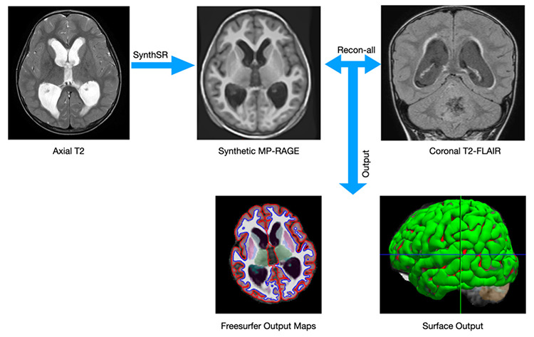
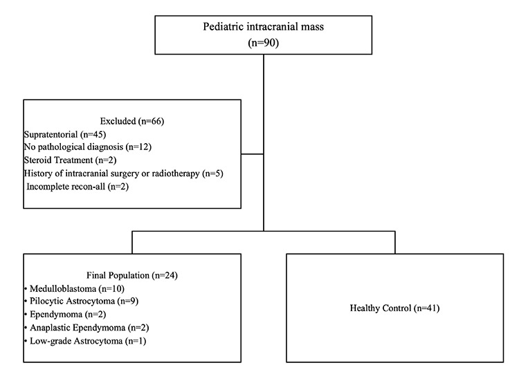
Methods: The study included 24 patients aged 4-18 years who were diagnosed with primary infratentorial tumors and 41 age- and gender-matched healthy controls. Synthetic magnetization-prepared rapid gradient echo images of brain magnetic resonance imaging were generated using deep learning algorithms applied to T2-axial images. The cortical thickness, surface area, volume, and local gyrification index (LGI), as well as subcortical gray matter volumes, were automatically calculated. Surface-based morphometry parameters for the patient and control groups were compared using the general linear model, and volumes between subcortical structures were compared using the t-test and Mann-Whitney U test.
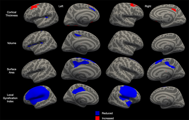
Results: In the patient group, cortical thinning was observed in the left supramarginal, and cortical thickening was observed in the left caudal middle frontal (CMF), left fusiform, left lateral orbitofrontal, left lingual gyrus, right CMF, right posterior cingulate, and right superior frontal (P < 0.050). The patient group showed a volume reduction in the pars triangularis, paracentral, precentral, and supramarginal gyri of the left hemisphere (P < 0.05). A decreased surface area was observed in the bilateral superior frontal and cingulate gyri (P < 0.05). The patient group exhibited a decreased LGI in the right precentral and superior temporal gyri, left supramarginal, and posterior cingulate gyri and showed an increased volume in the bilateral caudate nucleus and hippocampus, while a volume reduction was observed in the bilateral putamen, pallidum, and amygdala (P < 0.05). The ventricular volume and tumor volume showed a positive correlation with the cortical thickness in the bilateral CMF while demonstrating a negative correlation with areas exhibiting a decreased LGI (P < 0.05).
Conclusion: Posterior fossa tumors lead to widespread morphological changes in cortical structures, with the most prominent pattern being hypogyria.
Clinical significance: This study illuminates the neurological impacts of infratentorial tumors in children, providing a foundation for future therapeutic strategies aimed at mitigating these adverse cortical and subcortical changes and improving patient outcomes.
期刊介绍:
Diagnostic and Interventional Radiology (Diagn Interv Radiol) is the open access, online-only official publication of Turkish Society of Radiology. It is published bimonthly and the journal’s publication language is English.
The journal is a medium for original articles, reviews, pictorial essays, technical notes related to all fields of diagnostic and interventional radiology.


 分享
分享
 求助内容:
求助内容: 应助结果提醒方式:
 扫码关注我们
