{"title":"利用拉曼光谱对干细胞在二维和三维基底上的分化进行比较分析","authors":"F. Ravera, E. Efeoglu and H. J. Byrne","doi":"10.1039/D4AN00315B","DOIUrl":null,"url":null,"abstract":"<p >Chondrogenesis is a complex cellular process that involves the transformation of mesenchymal stem cells (MSCs) into chondrocytes, the specialised cells that form cartilage. In recent years, three-dimensional (3D) culture systems have emerged as a promising approach to studying cell behaviour and development in a more physiologically relevant environment compared to traditional two-dimensional (2D) cell culture. The use of these systems provided insights into the molecular mechanisms that regulate chondrogenesis and has the potential to revolutionise the development of new therapies for cartilage repair and regeneration. This study demonstrates the successful application of Raman microspectroscopy (RMS) as a label-free, non-destructive, and sensitive method to monitor the chondrogenic differentiation of bone marrow-derived rat mesenchymal stem cells (rMSCs) in a collagen type I hydrogel, and explores the potential benefits of 3D hydrogels compared to conventional 2D cell culture environments. rMSCs were cultured on 3D substrates for 3 weeks and their differentiation was monitored by measuring the spectral signatures of their subcellular compartments. Additionally, the evolution of high-density micromass cultures was investigated to provide a comprehensive understanding of the process and complex interactions between cells and their surrounding extracellular matrix. For comparison, rMSCs were induced into chondrogenesis in identical medium conditions for 21 days in monolayer culture. Raman spectra showed that rMSCs cultured in a collagen type I hydrogel are able to undergo a distinct chondrogenic differentiation pathway at a significantly higher rate than the 2D culture cells. 3D cultures expressed stronger and more homogeneous chondrogenesis-associated peaks such as collagens, glycosaminoglycans (GAGs), and aggrecan while manifesting changes in proteins and lipidic content. These results suggest that 3D type I collagen hydrogel substrates are promising for <em>in vitro</em> chondrogenesis studies, and that RMS is a valuable tool for monitoring chondrogenesis in 3D environments.</p>","PeriodicalId":63,"journal":{"name":"Analyst","volume":" 15","pages":" 4041-4053"},"PeriodicalIF":3.6000,"publicationDate":"2024-06-04","publicationTypes":"Journal Article","fieldsOfStudy":null,"isOpenAccess":false,"openAccessPdf":"","citationCount":"0","resultStr":"{\"title\":\"A comparative analysis of stem cell differentiation on 2D and 3D substrates using Raman microspectroscopy†\",\"authors\":\"F. Ravera, E. Efeoglu and H. J. Byrne\",\"doi\":\"10.1039/D4AN00315B\",\"DOIUrl\":null,\"url\":null,\"abstract\":\"<p >Chondrogenesis is a complex cellular process that involves the transformation of mesenchymal stem cells (MSCs) into chondrocytes, the specialised cells that form cartilage. In recent years, three-dimensional (3D) culture systems have emerged as a promising approach to studying cell behaviour and development in a more physiologically relevant environment compared to traditional two-dimensional (2D) cell culture. The use of these systems provided insights into the molecular mechanisms that regulate chondrogenesis and has the potential to revolutionise the development of new therapies for cartilage repair and regeneration. This study demonstrates the successful application of Raman microspectroscopy (RMS) as a label-free, non-destructive, and sensitive method to monitor the chondrogenic differentiation of bone marrow-derived rat mesenchymal stem cells (rMSCs) in a collagen type I hydrogel, and explores the potential benefits of 3D hydrogels compared to conventional 2D cell culture environments. rMSCs were cultured on 3D substrates for 3 weeks and their differentiation was monitored by measuring the spectral signatures of their subcellular compartments. Additionally, the evolution of high-density micromass cultures was investigated to provide a comprehensive understanding of the process and complex interactions between cells and their surrounding extracellular matrix. For comparison, rMSCs were induced into chondrogenesis in identical medium conditions for 21 days in monolayer culture. Raman spectra showed that rMSCs cultured in a collagen type I hydrogel are able to undergo a distinct chondrogenic differentiation pathway at a significantly higher rate than the 2D culture cells. 3D cultures expressed stronger and more homogeneous chondrogenesis-associated peaks such as collagens, glycosaminoglycans (GAGs), and aggrecan while manifesting changes in proteins and lipidic content. These results suggest that 3D type I collagen hydrogel substrates are promising for <em>in vitro</em> chondrogenesis studies, and that RMS is a valuable tool for monitoring chondrogenesis in 3D environments.</p>\",\"PeriodicalId\":63,\"journal\":{\"name\":\"Analyst\",\"volume\":\" 15\",\"pages\":\" 4041-4053\"},\"PeriodicalIF\":3.6000,\"publicationDate\":\"2024-06-04\",\"publicationTypes\":\"Journal Article\",\"fieldsOfStudy\":null,\"isOpenAccess\":false,\"openAccessPdf\":\"\",\"citationCount\":\"0\",\"resultStr\":null,\"platform\":\"Semanticscholar\",\"paperid\":null,\"PeriodicalName\":\"Analyst\",\"FirstCategoryId\":\"92\",\"ListUrlMain\":\"https://pubs.rsc.org/en/content/articlelanding/2024/an/d4an00315b\",\"RegionNum\":3,\"RegionCategory\":\"化学\",\"ArticlePicture\":[],\"TitleCN\":null,\"AbstractTextCN\":null,\"PMCID\":null,\"EPubDate\":\"\",\"PubModel\":\"\",\"JCR\":\"Q2\",\"JCRName\":\"CHEMISTRY, ANALYTICAL\",\"Score\":null,\"Total\":0}","platform":"Semanticscholar","paperid":null,"PeriodicalName":"Analyst","FirstCategoryId":"92","ListUrlMain":"https://pubs.rsc.org/en/content/articlelanding/2024/an/d4an00315b","RegionNum":3,"RegionCategory":"化学","ArticlePicture":[],"TitleCN":null,"AbstractTextCN":null,"PMCID":null,"EPubDate":"","PubModel":"","JCR":"Q2","JCRName":"CHEMISTRY, ANALYTICAL","Score":null,"Total":0}
引用次数: 0
摘要
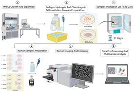
软骨形成是一个复杂的细胞过程,涉及间充质干细胞(MSCs)向软骨细胞(形成软骨的特化细胞)的转化。近年来,与传统的二维(2D)细胞培养相比,三维(3D)培养系统已成为在更贴近生理的环境中研究细胞行为和发育的一种有前途的方法。利用这些系统可以深入了解调控软骨生成的分子机制,并有可能彻底改变软骨修复和再生新疗法的开发。本研究展示了拉曼光谱(RMS)作为一种无标记、非破坏性和灵敏的方法在监测骨髓来源的大鼠间充质干细胞(rMSCs)在 I 型胶原水凝胶中的软骨分化方面的成功应用,并探索了三维水凝胶与传统二维细胞培养环境相比的潜在优势。rMSCs 在三维基底上培养了 3 周,通过测量其亚细胞区的光谱特征监测其分化。此外,还对高密度微质量培养物的演变进行了研究,以全面了解细胞与其周围细胞外基质之间的过程和复杂的相互作用。为了进行比较,在相同的培养基条件下对 rMSCs 进行了为期 21 天的单层培养,诱导其形成软骨。拉曼光谱显示,在 I 型胶原水凝胶中培养的 rMSCs 能够以明显高于二维培养细胞的速度经历独特的软骨分化途径。三维培养物表达的胶原蛋白、糖胺聚糖(GAG)和凝集素等软骨形成相关峰值更强、更均匀,同时蛋白质和脂质含量也发生了变化。这些结果表明,三维 I 型胶原水凝胶基底有望用于体外软骨形成研究,而 RMS 是监测三维环境中软骨形成的重要工具。
A comparative analysis of stem cell differentiation on 2D and 3D substrates using Raman microspectroscopy†
Chondrogenesis is a complex cellular process that involves the transformation of mesenchymal stem cells (MSCs) into chondrocytes, the specialised cells that form cartilage. In recent years, three-dimensional (3D) culture systems have emerged as a promising approach to studying cell behaviour and development in a more physiologically relevant environment compared to traditional two-dimensional (2D) cell culture. The use of these systems provided insights into the molecular mechanisms that regulate chondrogenesis and has the potential to revolutionise the development of new therapies for cartilage repair and regeneration. This study demonstrates the successful application of Raman microspectroscopy (RMS) as a label-free, non-destructive, and sensitive method to monitor the chondrogenic differentiation of bone marrow-derived rat mesenchymal stem cells (rMSCs) in a collagen type I hydrogel, and explores the potential benefits of 3D hydrogels compared to conventional 2D cell culture environments. rMSCs were cultured on 3D substrates for 3 weeks and their differentiation was monitored by measuring the spectral signatures of their subcellular compartments. Additionally, the evolution of high-density micromass cultures was investigated to provide a comprehensive understanding of the process and complex interactions between cells and their surrounding extracellular matrix. For comparison, rMSCs were induced into chondrogenesis in identical medium conditions for 21 days in monolayer culture. Raman spectra showed that rMSCs cultured in a collagen type I hydrogel are able to undergo a distinct chondrogenic differentiation pathway at a significantly higher rate than the 2D culture cells. 3D cultures expressed stronger and more homogeneous chondrogenesis-associated peaks such as collagens, glycosaminoglycans (GAGs), and aggrecan while manifesting changes in proteins and lipidic content. These results suggest that 3D type I collagen hydrogel substrates are promising for in vitro chondrogenesis studies, and that RMS is a valuable tool for monitoring chondrogenesis in 3D environments.


 分享
分享
 求助内容:
求助内容: 应助结果提醒方式:
 扫码关注我们
