Daniel Holl, Wing Fung Hau, Anais Julien, Shervin Banitalebi, Jannis Kalkitsas, Soniya Savant, Enric Llorens-Bobadilla, Yann Herault, Guillaume Pavlovic, Mahmood Amiry-Moghaddam, David Oliveira Dias, Christian Göritz
{"title":"基质成纤维细胞对小鼠创伤后纤维化的不同起源和区域依赖性贡献","authors":"Daniel Holl, Wing Fung Hau, Anais Julien, Shervin Banitalebi, Jannis Kalkitsas, Soniya Savant, Enric Llorens-Bobadilla, Yann Herault, Guillaume Pavlovic, Mahmood Amiry-Moghaddam, David Oliveira Dias, Christian Göritz","doi":"10.1038/s41593-024-01678-4","DOIUrl":null,"url":null,"abstract":"Fibrotic scar tissue formation occurs in humans and mice. The fibrotic scar impairs tissue regeneration and functional recovery. However, the origin of scar-forming fibroblasts is unclear. Here, we show that stromal fibroblasts forming the fibrotic scar derive from two populations of perivascular cells after spinal cord injury (SCI) in adult mice of both sexes. We anatomically and transcriptionally identify the two cell populations as pericytes and perivascular fibroblasts. Fibroblasts and pericytes are enriched in the white and gray matter regions of the spinal cord, respectively. Both cell populations are recruited in response to SCI and inflammation. However, their contribution to fibrotic scar tissue depends on the location of the lesion. Upon injury, pericytes and perivascular fibroblasts become activated and transcriptionally converge on the generation of stromal myofibroblasts. Our results show that pericytes and perivascular fibroblasts contribute to the fibrotic scar in a region-dependent manner. The origin and composition of stromal fibroblasts in the fibrotic CNS scar are unclear. Here, the authors demonstrate that pericytes and perivascular fibroblasts contribute to fibrotic scarring following spinal cord injury in mice in a region-dependent manner.","PeriodicalId":19076,"journal":{"name":"Nature neuroscience","volume":"27 7","pages":"1285-1298"},"PeriodicalIF":20.0000,"publicationDate":"2024-06-07","publicationTypes":"Journal Article","fieldsOfStudy":null,"isOpenAccess":false,"openAccessPdf":"https://www.nature.com/articles/s41593-024-01678-4.pdf","citationCount":"0","resultStr":"{\"title\":\"Distinct origin and region-dependent contribution of stromal fibroblasts to fibrosis following traumatic injury in mice\",\"authors\":\"Daniel Holl, Wing Fung Hau, Anais Julien, Shervin Banitalebi, Jannis Kalkitsas, Soniya Savant, Enric Llorens-Bobadilla, Yann Herault, Guillaume Pavlovic, Mahmood Amiry-Moghaddam, David Oliveira Dias, Christian Göritz\",\"doi\":\"10.1038/s41593-024-01678-4\",\"DOIUrl\":null,\"url\":null,\"abstract\":\"Fibrotic scar tissue formation occurs in humans and mice. The fibrotic scar impairs tissue regeneration and functional recovery. However, the origin of scar-forming fibroblasts is unclear. Here, we show that stromal fibroblasts forming the fibrotic scar derive from two populations of perivascular cells after spinal cord injury (SCI) in adult mice of both sexes. We anatomically and transcriptionally identify the two cell populations as pericytes and perivascular fibroblasts. Fibroblasts and pericytes are enriched in the white and gray matter regions of the spinal cord, respectively. Both cell populations are recruited in response to SCI and inflammation. However, their contribution to fibrotic scar tissue depends on the location of the lesion. Upon injury, pericytes and perivascular fibroblasts become activated and transcriptionally converge on the generation of stromal myofibroblasts. Our results show that pericytes and perivascular fibroblasts contribute to the fibrotic scar in a region-dependent manner. The origin and composition of stromal fibroblasts in the fibrotic CNS scar are unclear. Here, the authors demonstrate that pericytes and perivascular fibroblasts contribute to fibrotic scarring following spinal cord injury in mice in a region-dependent manner.\",\"PeriodicalId\":19076,\"journal\":{\"name\":\"Nature neuroscience\",\"volume\":\"27 7\",\"pages\":\"1285-1298\"},\"PeriodicalIF\":20.0000,\"publicationDate\":\"2024-06-07\",\"publicationTypes\":\"Journal Article\",\"fieldsOfStudy\":null,\"isOpenAccess\":false,\"openAccessPdf\":\"https://www.nature.com/articles/s41593-024-01678-4.pdf\",\"citationCount\":\"0\",\"resultStr\":null,\"platform\":\"Semanticscholar\",\"paperid\":null,\"PeriodicalName\":\"Nature neuroscience\",\"FirstCategoryId\":\"3\",\"ListUrlMain\":\"https://www.nature.com/articles/s41593-024-01678-4\",\"RegionNum\":1,\"RegionCategory\":\"医学\",\"ArticlePicture\":[],\"TitleCN\":null,\"AbstractTextCN\":null,\"PMCID\":null,\"EPubDate\":\"\",\"PubModel\":\"\",\"JCR\":\"Q1\",\"JCRName\":\"NEUROSCIENCES\",\"Score\":null,\"Total\":0}","platform":"Semanticscholar","paperid":null,"PeriodicalName":"Nature neuroscience","FirstCategoryId":"3","ListUrlMain":"https://www.nature.com/articles/s41593-024-01678-4","RegionNum":1,"RegionCategory":"医学","ArticlePicture":[],"TitleCN":null,"AbstractTextCN":null,"PMCID":null,"EPubDate":"","PubModel":"","JCR":"Q1","JCRName":"NEUROSCIENCES","Score":null,"Total":0}
Distinct origin and region-dependent contribution of stromal fibroblasts to fibrosis following traumatic injury in mice
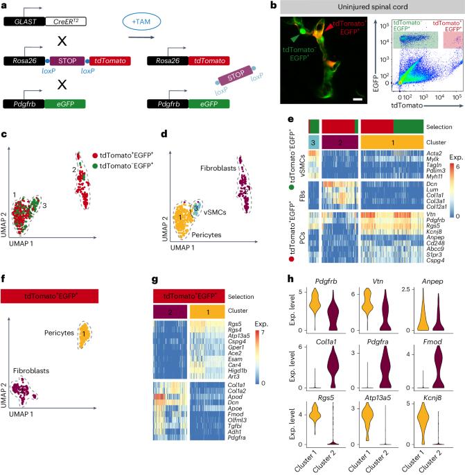
Fibrotic scar tissue formation occurs in humans and mice. The fibrotic scar impairs tissue regeneration and functional recovery. However, the origin of scar-forming fibroblasts is unclear. Here, we show that stromal fibroblasts forming the fibrotic scar derive from two populations of perivascular cells after spinal cord injury (SCI) in adult mice of both sexes. We anatomically and transcriptionally identify the two cell populations as pericytes and perivascular fibroblasts. Fibroblasts and pericytes are enriched in the white and gray matter regions of the spinal cord, respectively. Both cell populations are recruited in response to SCI and inflammation. However, their contribution to fibrotic scar tissue depends on the location of the lesion. Upon injury, pericytes and perivascular fibroblasts become activated and transcriptionally converge on the generation of stromal myofibroblasts. Our results show that pericytes and perivascular fibroblasts contribute to the fibrotic scar in a region-dependent manner. The origin and composition of stromal fibroblasts in the fibrotic CNS scar are unclear. Here, the authors demonstrate that pericytes and perivascular fibroblasts contribute to fibrotic scarring following spinal cord injury in mice in a region-dependent manner.
期刊介绍:
Nature Neuroscience, a multidisciplinary journal, publishes papers of the utmost quality and significance across all realms of neuroscience. The editors welcome contributions spanning molecular, cellular, systems, and cognitive neuroscience, along with psychophysics, computational modeling, and nervous system disorders. While no area is off-limits, studies offering fundamental insights into nervous system function receive priority.
The journal offers high visibility to both readers and authors, fostering interdisciplinary communication and accessibility to a broad audience. It maintains high standards of copy editing and production, rigorous peer review, rapid publication, and operates independently from academic societies and other vested interests.
In addition to primary research, Nature Neuroscience features news and views, reviews, editorials, commentaries, perspectives, book reviews, and correspondence, aiming to serve as the voice of the global neuroscience community.


 分享
分享
 求助内容:
求助内容: 应助结果提醒方式:
 扫码关注我们
