Zhengteng Li, Hongmei Liu, Min Wang, Xiankai Wang, Dongmei Pan, Aidong Ma, Yang Chen
{"title":"基于临床和多层螺旋计算机断层扫描特征的IA期肺腺癌术前预测Ki-67表达和预后的提名图","authors":"Zhengteng Li, Hongmei Liu, Min Wang, Xiankai Wang, Dongmei Pan, Aidong Ma, Yang Chen","doi":"10.1186/s12880-024-01305-5","DOIUrl":null,"url":null,"abstract":"<p><strong>Objective: </strong>This study developed and validated a nomogram utilizing clinical and multi-slice spiral computed tomography (MSCT) features for the preoperative prediction of Ki-67 expression in stage IA lung adenocarcinoma. Additionally, we assessed the predictive accuracy of Ki-67 expression levels, as determined by our model, in estimating the prognosis of stage IA lung adenocarcinoma.</p><p><strong>Materials and methods: </strong>We retrospectively analyzed data from 395 patients with pathologically confirmed stage IA lung adenocarcinoma. A total of 322 patients were divided into training and internal validation groups at a 6:4 ratio, whereas the remaining 73 patients composed the external validation group. According to the pathological results, the patients were classified into high and low Ki-67 labeling index (LI) groups. Clinical and CT features were subjected to statistical analysis. The training group was used to construct a predictive model through logistic regression and to formulate a nomogram. The nomogram's predictive ability and goodness-of-fit were assessed. Internal and external validations were performed, and clinical utility was evaluated. Finally, the recurrence-free survival (RFS) rates were compared.</p><p><strong>Results: </strong>In the training group, sex, age, tumor density type, tumor-lung interface, lobulation, spiculation, pleural indentation, and maximum nodule diameter differed significantly between patients with high and low Ki-67 LI. Multivariate logistic regression analysis revealed that sex, tumor density, and maximum nodule diameter were significantly associated with high Ki-67 expression in stage IA lung adenocarcinoma. The calibration curves closely resembled the standard curves, indicating the excellent discrimination and accuracy of the model. Decision curve analysis revealed favorable clinical utility. Patients with a nomogram-predicted high Ki-67 LI exhibited worse RFS.</p><p><strong>Conclusion: </strong>The nomogram utilizing clinical and CT features for the preoperative prediction of Ki-67 expression in stage IA lung adenocarcinoma demonstrated excellent performance, clinical utility, and prognostic significance, suggesting that this nomogram is a noninvasive personalized approach for the preoperative prediction of Ki-67 expression.</p>","PeriodicalId":9020,"journal":{"name":"BMC Medical Imaging","volume":"24 1","pages":"143"},"PeriodicalIF":3.2000,"publicationDate":"2024-06-12","publicationTypes":"Journal Article","fieldsOfStudy":null,"isOpenAccess":false,"openAccessPdf":"https://www.ncbi.nlm.nih.gov/pmc/articles/PMC11167796/pdf/","citationCount":"0","resultStr":"{\"title\":\"Nomogram for the preoperative prediction of Ki-67 expression and prognosis in stage IA lung adenocarcinoma based on clinical and multi-slice spiral computed tomography features.\",\"authors\":\"Zhengteng Li, Hongmei Liu, Min Wang, Xiankai Wang, Dongmei Pan, Aidong Ma, Yang Chen\",\"doi\":\"10.1186/s12880-024-01305-5\",\"DOIUrl\":null,\"url\":null,\"abstract\":\"<p><strong>Objective: </strong>This study developed and validated a nomogram utilizing clinical and multi-slice spiral computed tomography (MSCT) features for the preoperative prediction of Ki-67 expression in stage IA lung adenocarcinoma. Additionally, we assessed the predictive accuracy of Ki-67 expression levels, as determined by our model, in estimating the prognosis of stage IA lung adenocarcinoma.</p><p><strong>Materials and methods: </strong>We retrospectively analyzed data from 395 patients with pathologically confirmed stage IA lung adenocarcinoma. A total of 322 patients were divided into training and internal validation groups at a 6:4 ratio, whereas the remaining 73 patients composed the external validation group. According to the pathological results, the patients were classified into high and low Ki-67 labeling index (LI) groups. Clinical and CT features were subjected to statistical analysis. The training group was used to construct a predictive model through logistic regression and to formulate a nomogram. The nomogram's predictive ability and goodness-of-fit were assessed. Internal and external validations were performed, and clinical utility was evaluated. Finally, the recurrence-free survival (RFS) rates were compared.</p><p><strong>Results: </strong>In the training group, sex, age, tumor density type, tumor-lung interface, lobulation, spiculation, pleural indentation, and maximum nodule diameter differed significantly between patients with high and low Ki-67 LI. Multivariate logistic regression analysis revealed that sex, tumor density, and maximum nodule diameter were significantly associated with high Ki-67 expression in stage IA lung adenocarcinoma. The calibration curves closely resembled the standard curves, indicating the excellent discrimination and accuracy of the model. Decision curve analysis revealed favorable clinical utility. Patients with a nomogram-predicted high Ki-67 LI exhibited worse RFS.</p><p><strong>Conclusion: </strong>The nomogram utilizing clinical and CT features for the preoperative prediction of Ki-67 expression in stage IA lung adenocarcinoma demonstrated excellent performance, clinical utility, and prognostic significance, suggesting that this nomogram is a noninvasive personalized approach for the preoperative prediction of Ki-67 expression.</p>\",\"PeriodicalId\":9020,\"journal\":{\"name\":\"BMC Medical Imaging\",\"volume\":\"24 1\",\"pages\":\"143\"},\"PeriodicalIF\":3.2000,\"publicationDate\":\"2024-06-12\",\"publicationTypes\":\"Journal Article\",\"fieldsOfStudy\":null,\"isOpenAccess\":false,\"openAccessPdf\":\"https://www.ncbi.nlm.nih.gov/pmc/articles/PMC11167796/pdf/\",\"citationCount\":\"0\",\"resultStr\":null,\"platform\":\"Semanticscholar\",\"paperid\":null,\"PeriodicalName\":\"BMC Medical Imaging\",\"FirstCategoryId\":\"3\",\"ListUrlMain\":\"https://doi.org/10.1186/s12880-024-01305-5\",\"RegionNum\":3,\"RegionCategory\":\"医学\",\"ArticlePicture\":[],\"TitleCN\":null,\"AbstractTextCN\":null,\"PMCID\":null,\"EPubDate\":\"\",\"PubModel\":\"\",\"JCR\":\"Q2\",\"JCRName\":\"RADIOLOGY, NUCLEAR MEDICINE & MEDICAL IMAGING\",\"Score\":null,\"Total\":0}","platform":"Semanticscholar","paperid":null,"PeriodicalName":"BMC Medical Imaging","FirstCategoryId":"3","ListUrlMain":"https://doi.org/10.1186/s12880-024-01305-5","RegionNum":3,"RegionCategory":"医学","ArticlePicture":[],"TitleCN":null,"AbstractTextCN":null,"PMCID":null,"EPubDate":"","PubModel":"","JCR":"Q2","JCRName":"RADIOLOGY, NUCLEAR MEDICINE & MEDICAL IMAGING","Score":null,"Total":0}
引用次数: 0
摘要
研究目的本研究利用临床和多层螺旋计算机断层扫描(MSCT)特征开发并验证了一种用于术前预测IA期肺腺癌Ki-67表达的提名图。此外,我们还评估了由我们的模型确定的 Ki-67 表达水平在估计 IA 期肺腺癌预后方面的预测准确性:我们回顾性分析了395例经病理证实的IA期肺腺癌患者的数据。共有 322 名患者按 6:4 的比例被分为训练组和内部验证组,其余 73 名患者组成外部验证组。根据病理结果,患者被分为高Ki-67标记指数(LI)组和低Ki-67标记指数(LI)组。对临床和 CT 特征进行统计分析。训练组通过逻辑回归构建了预测模型,并制定了提名图。对提名图的预测能力和拟合优度进行了评估。进行了内部和外部验证,并评估了临床实用性。最后,比较了无复发生存率(RFS):在训练组中,Ki-67 LI 高和 Ki-67 LI 低的患者在性别、年龄、肿瘤密度类型、肿瘤-肺界面、分叶、棘点、胸膜压痕和结节最大直径方面存在显著差异。多变量逻辑回归分析显示,性别、肿瘤密度和最大结节直径与IA期肺腺癌的Ki-67高表达有明显相关性。校准曲线与标准曲线非常相似,表明该模型具有极佳的区分度和准确性。决策曲线分析显示了良好的临床实用性。提名图预测的高Ki-67 LI患者的RFS较差:结论:利用临床和CT特征进行IA期肺腺癌术前Ki-67表达预测的提名图表现出了卓越的性能、临床实用性和预后意义,表明该提名图是一种无创的个性化Ki-67表达术前预测方法。
Nomogram for the preoperative prediction of Ki-67 expression and prognosis in stage IA lung adenocarcinoma based on clinical and multi-slice spiral computed tomography features.
Objective: This study developed and validated a nomogram utilizing clinical and multi-slice spiral computed tomography (MSCT) features for the preoperative prediction of Ki-67 expression in stage IA lung adenocarcinoma. Additionally, we assessed the predictive accuracy of Ki-67 expression levels, as determined by our model, in estimating the prognosis of stage IA lung adenocarcinoma.
Materials and methods: We retrospectively analyzed data from 395 patients with pathologically confirmed stage IA lung adenocarcinoma. A total of 322 patients were divided into training and internal validation groups at a 6:4 ratio, whereas the remaining 73 patients composed the external validation group. According to the pathological results, the patients were classified into high and low Ki-67 labeling index (LI) groups. Clinical and CT features were subjected to statistical analysis. The training group was used to construct a predictive model through logistic regression and to formulate a nomogram. The nomogram's predictive ability and goodness-of-fit were assessed. Internal and external validations were performed, and clinical utility was evaluated. Finally, the recurrence-free survival (RFS) rates were compared.
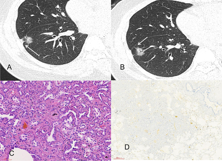
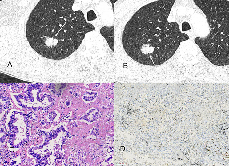
Results: In the training group, sex, age, tumor density type, tumor-lung interface, lobulation, spiculation, pleural indentation, and maximum nodule diameter differed significantly between patients with high and low Ki-67 LI. Multivariate logistic regression analysis revealed that sex, tumor density, and maximum nodule diameter were significantly associated with high Ki-67 expression in stage IA lung adenocarcinoma. The calibration curves closely resembled the standard curves, indicating the excellent discrimination and accuracy of the model. Decision curve analysis revealed favorable clinical utility. Patients with a nomogram-predicted high Ki-67 LI exhibited worse RFS.
Conclusion: The nomogram utilizing clinical and CT features for the preoperative prediction of Ki-67 expression in stage IA lung adenocarcinoma demonstrated excellent performance, clinical utility, and prognostic significance, suggesting that this nomogram is a noninvasive personalized approach for the preoperative prediction of Ki-67 expression.
期刊介绍:
BMC Medical Imaging is an open access journal publishing original peer-reviewed research articles in the development, evaluation, and use of imaging techniques and image processing tools to diagnose and manage disease.


 分享
分享
 求助内容:
求助内容: 应助结果提醒方式:
 扫码关注我们
