{"title":"s2MRI-ADNet:一种可解释的深度学习框架,仅从结构性核磁共振成像中整合阿尔茨海默病的欧氏图表征。","authors":"Zhiwei Song, Honglun Li, Yiyu Zhang, Chuanzhen Zhu, Minbo Jiang, Limei Song, Yi Wang, Minhui Ouyang, Fang Hu, Qiang Zheng","doi":"10.1007/s10334-024-01178-3","DOIUrl":null,"url":null,"abstract":"<p><strong>Objective: </strong>To establish a multi-dimensional representation solely on structural MRI (sMRI) for early diagnosis of AD.</p><p><strong>Methods: </strong>A total of 3377 participants' sMRI from four independent databases were retrospectively identified to construct an interpretable deep learning model that integrated multi-dimensional representations of AD solely on sMRI (called s<sup>2</sup>MRI-ADNet) by a dual-channel learning strategy of gray matter volume (GMV) from Euclidean space and the regional radiomics similarity network (R2SN) from graph space. Specifically, the GMV feature map learning channel (called GMV-Channel) was to take into consideration spatial information of both long-range spatial relations and detailed localization information, while the node feature and connectivity strength learning channel (called NFCS-Channel) was to characterize the graph-structured R2SN network by a separable learning strategy.</p><p><strong>Results: </strong>The s<sup>2</sup>MRI-ADNet achieved a superior classification accuracy of 92.1% and 91.4% under intra-database and inter-database cross-validation. The GMV-Channel and NFCS-Channel captured complementary group-discriminative brain regions, revealing a complementary interpretation of the multi-dimensional representation of brain structure in Euclidean and graph spaces respectively. Besides, the generalizable and reproducible interpretation of the multi-dimensional representation in capturing complementary group-discriminative brain regions revealed a significant correlation between the four independent databases (p < 0.05). Significant associations (p < 0.05) between attention scores and brain abnormality, between classification scores and clinical measure of cognitive ability, CSF biomarker, metabolism, and genetic risk score also provided solid neurobiological interpretation.</p><p><strong>Conclusion: </strong>The s<sup>2</sup>MRI-ADNet solely on sMRI could leverage the complementary multi-dimensional representations of AD in Euclidean and graph spaces, and achieved superior performance in the early diagnosis of AD, facilitating its potential in both clinical translation and popularization.</p>","PeriodicalId":18067,"journal":{"name":"Magnetic Resonance Materials in Physics, Biology and Medicine","volume":" ","pages":"845-857"},"PeriodicalIF":3.1000,"publicationDate":"2024-10-01","publicationTypes":"Journal Article","fieldsOfStudy":null,"isOpenAccess":false,"openAccessPdf":"","citationCount":"0","resultStr":"{\"title\":\"s<sup>2</sup>MRI-ADNet: an interpretable deep learning framework integrating Euclidean-graph representations of Alzheimer's disease solely from structural MRI.\",\"authors\":\"Zhiwei Song, Honglun Li, Yiyu Zhang, Chuanzhen Zhu, Minbo Jiang, Limei Song, Yi Wang, Minhui Ouyang, Fang Hu, Qiang Zheng\",\"doi\":\"10.1007/s10334-024-01178-3\",\"DOIUrl\":null,\"url\":null,\"abstract\":\"<p><strong>Objective: </strong>To establish a multi-dimensional representation solely on structural MRI (sMRI) for early diagnosis of AD.</p><p><strong>Methods: </strong>A total of 3377 participants' sMRI from four independent databases were retrospectively identified to construct an interpretable deep learning model that integrated multi-dimensional representations of AD solely on sMRI (called s<sup>2</sup>MRI-ADNet) by a dual-channel learning strategy of gray matter volume (GMV) from Euclidean space and the regional radiomics similarity network (R2SN) from graph space. Specifically, the GMV feature map learning channel (called GMV-Channel) was to take into consideration spatial information of both long-range spatial relations and detailed localization information, while the node feature and connectivity strength learning channel (called NFCS-Channel) was to characterize the graph-structured R2SN network by a separable learning strategy.</p><p><strong>Results: </strong>The s<sup>2</sup>MRI-ADNet achieved a superior classification accuracy of 92.1% and 91.4% under intra-database and inter-database cross-validation. The GMV-Channel and NFCS-Channel captured complementary group-discriminative brain regions, revealing a complementary interpretation of the multi-dimensional representation of brain structure in Euclidean and graph spaces respectively. Besides, the generalizable and reproducible interpretation of the multi-dimensional representation in capturing complementary group-discriminative brain regions revealed a significant correlation between the four independent databases (p < 0.05). Significant associations (p < 0.05) between attention scores and brain abnormality, between classification scores and clinical measure of cognitive ability, CSF biomarker, metabolism, and genetic risk score also provided solid neurobiological interpretation.</p><p><strong>Conclusion: </strong>The s<sup>2</sup>MRI-ADNet solely on sMRI could leverage the complementary multi-dimensional representations of AD in Euclidean and graph spaces, and achieved superior performance in the early diagnosis of AD, facilitating its potential in both clinical translation and popularization.</p>\",\"PeriodicalId\":18067,\"journal\":{\"name\":\"Magnetic Resonance Materials in Physics, Biology and Medicine\",\"volume\":\" \",\"pages\":\"845-857\"},\"PeriodicalIF\":3.1000,\"publicationDate\":\"2024-10-01\",\"publicationTypes\":\"Journal Article\",\"fieldsOfStudy\":null,\"isOpenAccess\":false,\"openAccessPdf\":\"\",\"citationCount\":\"0\",\"resultStr\":null,\"platform\":\"Semanticscholar\",\"paperid\":null,\"PeriodicalName\":\"Magnetic Resonance Materials in Physics, Biology and Medicine\",\"FirstCategoryId\":\"3\",\"ListUrlMain\":\"https://doi.org/10.1007/s10334-024-01178-3\",\"RegionNum\":4,\"RegionCategory\":\"医学\",\"ArticlePicture\":[],\"TitleCN\":null,\"AbstractTextCN\":null,\"PMCID\":null,\"EPubDate\":\"2024/6/13 0:00:00\",\"PubModel\":\"Epub\",\"JCR\":\"Q3\",\"JCRName\":\"RADIOLOGY, NUCLEAR MEDICINE & MEDICAL IMAGING\",\"Score\":null,\"Total\":0}","platform":"Semanticscholar","paperid":null,"PeriodicalName":"Magnetic Resonance Materials in Physics, Biology and Medicine","FirstCategoryId":"3","ListUrlMain":"https://doi.org/10.1007/s10334-024-01178-3","RegionNum":4,"RegionCategory":"医学","ArticlePicture":[],"TitleCN":null,"AbstractTextCN":null,"PMCID":null,"EPubDate":"2024/6/13 0:00:00","PubModel":"Epub","JCR":"Q3","JCRName":"RADIOLOGY, NUCLEAR MEDICINE & MEDICAL IMAGING","Score":null,"Total":0}
引用次数: 0
摘要
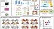
目的方法:从四个独立的数据库中回顾性地识别了3377名参与者的sMRI,并构建了一个可解释的深度学习模型,该模型仅在结构性磁共振成像(sMRI)上整合了AD的多维表征,用于早期诊断AD:方法:回顾性鉴定了四个独立数据库中3377名参与者的sMRI,通过欧几里得空间的灰质体积(GMV)和图空间的区域放射组学相似性网络(R2SN)的双通道学习策略,构建了一个可解释的深度学习模型,该模型仅在sMRI上整合了AD的多维表征(称为s2MRI-ADNet)。具体来说,GMV特征图学习通道(称为GMV通道)考虑了长程空间关系的空间信息和详细的定位信息,而节点特征和连接强度学习通道(称为NFCS通道)则通过可分离的学习策略来表征图结构的R2SN网络:结果:s2MRI-ADNet 在数据库内和数据库间交叉验证中的分类准确率分别达到 92.1% 和 91.4%。GMV通道和NFCS通道捕捉到了互补的分组区分脑区,分别揭示了欧几里得空间和图空间中脑结构多维表征的互补性解释。此外,在捕捉互补性组别区分脑区方面,对多维表征的解释具有普遍性和可重复性,这揭示了四个独立数据库之间的显著相关性(p 结论):仅基于 sMRI 的 s2MRI-ADNet 可利用欧几里得空间和图空间中互补的 AD 多维表征,在 AD 早期诊断方面取得了优异的表现,促进了其在临床转化和推广方面的潜力。
s2MRI-ADNet: an interpretable deep learning framework integrating Euclidean-graph representations of Alzheimer's disease solely from structural MRI.
Objective: To establish a multi-dimensional representation solely on structural MRI (sMRI) for early diagnosis of AD.
Methods: A total of 3377 participants' sMRI from four independent databases were retrospectively identified to construct an interpretable deep learning model that integrated multi-dimensional representations of AD solely on sMRI (called s2MRI-ADNet) by a dual-channel learning strategy of gray matter volume (GMV) from Euclidean space and the regional radiomics similarity network (R2SN) from graph space. Specifically, the GMV feature map learning channel (called GMV-Channel) was to take into consideration spatial information of both long-range spatial relations and detailed localization information, while the node feature and connectivity strength learning channel (called NFCS-Channel) was to characterize the graph-structured R2SN network by a separable learning strategy.
Results: The s2MRI-ADNet achieved a superior classification accuracy of 92.1% and 91.4% under intra-database and inter-database cross-validation. The GMV-Channel and NFCS-Channel captured complementary group-discriminative brain regions, revealing a complementary interpretation of the multi-dimensional representation of brain structure in Euclidean and graph spaces respectively. Besides, the generalizable and reproducible interpretation of the multi-dimensional representation in capturing complementary group-discriminative brain regions revealed a significant correlation between the four independent databases (p < 0.05). Significant associations (p < 0.05) between attention scores and brain abnormality, between classification scores and clinical measure of cognitive ability, CSF biomarker, metabolism, and genetic risk score also provided solid neurobiological interpretation.
Conclusion: The s2MRI-ADNet solely on sMRI could leverage the complementary multi-dimensional representations of AD in Euclidean and graph spaces, and achieved superior performance in the early diagnosis of AD, facilitating its potential in both clinical translation and popularization.
期刊介绍:
MAGMA is a multidisciplinary international journal devoted to the publication of articles on all aspects of magnetic resonance techniques and their applications in medicine and biology. MAGMA currently publishes research papers, reviews, letters to the editor, and commentaries, six times a year. The subject areas covered by MAGMA include:
advances in materials, hardware and software in magnetic resonance technology,
new developments and results in research and practical applications of magnetic resonance imaging and spectroscopy related to biology and medicine,
study of animal models and intact cells using magnetic resonance,
reports of clinical trials on humans and clinical validation of magnetic resonance protocols.


 分享
分享
 求助内容:
求助内容: 应助结果提醒方式:
 扫码关注我们
