Pingwei Xu, Junjie Chi, Xiaochen Wang, Meng Zhu, Kai Chen, Qihui Fan, Fangfu Ye and Changmin Shao
{"title":"基于微流体反蛋白石支架的体外血管化肝脏肿瘤模型,用于免疫细胞招募研究。","authors":"Pingwei Xu, Junjie Chi, Xiaochen Wang, Meng Zhu, Kai Chen, Qihui Fan, Fangfu Ye and Changmin Shao","doi":"10.1039/D4LC00341A","DOIUrl":null,"url":null,"abstract":"<p >Liver cancer, characterized as a kind of malignant tumor within the digestive system, poses great health harm, and immune escape stands out as an important reason for its occurrence and development. Chemokines, pivotal in guiding immune cells' migration, is necessary to initiate and deliver an effective anti-tumor immune response. Therefore, understanding the chemotactic environment and identifying chemokines that regulate recruitment of immune cells to the tumor microenvironment (TME) are critical to improve current immunotherapy interventions. Herein, we report a well-defined inverse opal scaffold generated with a microfluidic emulsion template for the construction of a vascularized liver tumor model, offering insights into immune cells' recruitment. Due to the excellent 3D porous morphology of the inverse opal scaffold, human hepatocellular carcinoma cells can aggregate in the pores of the scaffold to form uniform multicellular tumor spheroids. More attractively, the vascularized liver tumor model can be achieved by constructing a 3D co-culture system involving endothelial cells and hepatocellular carcinoma cells. The results demonstrate that the 3D co-cultured tumor cells increase the neutrophil chemokines remarkably and recruit neutrophils to tumor tissues, then promote tumor progression. This approach opens a feasible avenue for realizing a vascularized liver tumor model with a reliable immune microenvironment close to that of a solid tumor of liver cancer.</p>","PeriodicalId":85,"journal":{"name":"Lab on a Chip","volume":" 14","pages":" 3470-3479"},"PeriodicalIF":6.3000,"publicationDate":"2024-06-19","publicationTypes":"Journal Article","fieldsOfStudy":null,"isOpenAccess":false,"openAccessPdf":"","citationCount":"0","resultStr":"{\"title\":\"In vitro vascularized liver tumor model based on a microfluidic inverse opal scaffold for immune cell recruitment investigation†\",\"authors\":\"Pingwei Xu, Junjie Chi, Xiaochen Wang, Meng Zhu, Kai Chen, Qihui Fan, Fangfu Ye and Changmin Shao\",\"doi\":\"10.1039/D4LC00341A\",\"DOIUrl\":null,\"url\":null,\"abstract\":\"<p >Liver cancer, characterized as a kind of malignant tumor within the digestive system, poses great health harm, and immune escape stands out as an important reason for its occurrence and development. Chemokines, pivotal in guiding immune cells' migration, is necessary to initiate and deliver an effective anti-tumor immune response. Therefore, understanding the chemotactic environment and identifying chemokines that regulate recruitment of immune cells to the tumor microenvironment (TME) are critical to improve current immunotherapy interventions. Herein, we report a well-defined inverse opal scaffold generated with a microfluidic emulsion template for the construction of a vascularized liver tumor model, offering insights into immune cells' recruitment. Due to the excellent 3D porous morphology of the inverse opal scaffold, human hepatocellular carcinoma cells can aggregate in the pores of the scaffold to form uniform multicellular tumor spheroids. More attractively, the vascularized liver tumor model can be achieved by constructing a 3D co-culture system involving endothelial cells and hepatocellular carcinoma cells. The results demonstrate that the 3D co-cultured tumor cells increase the neutrophil chemokines remarkably and recruit neutrophils to tumor tissues, then promote tumor progression. This approach opens a feasible avenue for realizing a vascularized liver tumor model with a reliable immune microenvironment close to that of a solid tumor of liver cancer.</p>\",\"PeriodicalId\":85,\"journal\":{\"name\":\"Lab on a Chip\",\"volume\":\" 14\",\"pages\":\" 3470-3479\"},\"PeriodicalIF\":6.3000,\"publicationDate\":\"2024-06-19\",\"publicationTypes\":\"Journal Article\",\"fieldsOfStudy\":null,\"isOpenAccess\":false,\"openAccessPdf\":\"\",\"citationCount\":\"0\",\"resultStr\":null,\"platform\":\"Semanticscholar\",\"paperid\":null,\"PeriodicalName\":\"Lab on a Chip\",\"FirstCategoryId\":\"5\",\"ListUrlMain\":\"https://pubs.rsc.org/en/content/articlelanding/2024/lc/d4lc00341a\",\"RegionNum\":2,\"RegionCategory\":\"工程技术\",\"ArticlePicture\":[],\"TitleCN\":null,\"AbstractTextCN\":null,\"PMCID\":null,\"EPubDate\":\"\",\"PubModel\":\"\",\"JCR\":\"Q1\",\"JCRName\":\"BIOCHEMICAL RESEARCH METHODS\",\"Score\":null,\"Total\":0}","platform":"Semanticscholar","paperid":null,"PeriodicalName":"Lab on a Chip","FirstCategoryId":"5","ListUrlMain":"https://pubs.rsc.org/en/content/articlelanding/2024/lc/d4lc00341a","RegionNum":2,"RegionCategory":"工程技术","ArticlePicture":[],"TitleCN":null,"AbstractTextCN":null,"PMCID":null,"EPubDate":"","PubModel":"","JCR":"Q1","JCRName":"BIOCHEMICAL RESEARCH METHODS","Score":null,"Total":0}
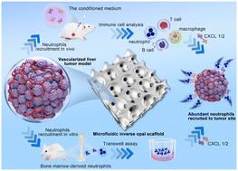
In vitro vascularized liver tumor model based on a microfluidic inverse opal scaffold for immune cell recruitment investigation†
Liver cancer, characterized as a kind of malignant tumor within the digestive system, poses great health harm, and immune escape stands out as an important reason for its occurrence and development. Chemokines, pivotal in guiding immune cells' migration, is necessary to initiate and deliver an effective anti-tumor immune response. Therefore, understanding the chemotactic environment and identifying chemokines that regulate recruitment of immune cells to the tumor microenvironment (TME) are critical to improve current immunotherapy interventions. Herein, we report a well-defined inverse opal scaffold generated with a microfluidic emulsion template for the construction of a vascularized liver tumor model, offering insights into immune cells' recruitment. Due to the excellent 3D porous morphology of the inverse opal scaffold, human hepatocellular carcinoma cells can aggregate in the pores of the scaffold to form uniform multicellular tumor spheroids. More attractively, the vascularized liver tumor model can be achieved by constructing a 3D co-culture system involving endothelial cells and hepatocellular carcinoma cells. The results demonstrate that the 3D co-cultured tumor cells increase the neutrophil chemokines remarkably and recruit neutrophils to tumor tissues, then promote tumor progression. This approach opens a feasible avenue for realizing a vascularized liver tumor model with a reliable immune microenvironment close to that of a solid tumor of liver cancer.
期刊介绍:
Lab on a Chip is the premiere journal that publishes cutting-edge research in the field of miniaturization. By their very nature, microfluidic/nanofluidic/miniaturized systems are at the intersection of disciplines, spanning fundamental research to high-end application, which is reflected by the broad readership of the journal. Lab on a Chip publishes two types of papers on original research: full-length research papers and communications. Papers should demonstrate innovations, which can come from technical advancements or applications addressing pressing needs in globally important areas. The journal also publishes Comments, Reviews, and Perspectives.


 分享
分享
 求助内容:
求助内容: 应助结果提醒方式:
 扫码关注我们
