{"title":"FAM172A 基因缺失可促进肝脏再生过程中的细胞增殖。","authors":"Herui Wei, Yifan Zhang, Meixin Gao, Junru Yang, Shiwei Wang, Xingang Zhou, Hongshan Wei, Fan Xiao","doi":"10.1007/s11010-024-05044-7","DOIUrl":null,"url":null,"abstract":"<p><p>The present study was designed to explore the function of FAM172A in liver regeneration and HCC. Mice were sacrificed after 70% partial hepatectomy (PH). RNA sequencing was performed on primary hepatocytes of WT and FAM172A<sup>-/-</sup> mice. We used HepG2 cells to construct cell lines with stably knockdown and overexpression of FAM172A. The expression of FAM172A in liver tissues was investigated by immunohistochemical staining, and we also used public database to perform survival analysis and prognostic model in HCC. Compared with WT mice after PH, normalized liver weight/body weight (LW/BW) ratio and the proliferating cell nuclear antigen (PCNA) protein level of FAM172A<sup>-/-</sup> mice elevated. The DEGs were mainly enriched in inflammatory response, tumor necrosis factor production, and wound healing. FAM172A knockdown enhanced the NFκB-TNFα and pERK-YAP1-Cyclin D1 axis. FAM172A peptide inhibited proliferation of primary hepatocytes. Moreover, the low expression of FAM172A in human HCC tissues implies a lower likelihood of survival and a valid diagnostic marker for HCC. Loss of FAM172A gene promotes cell proliferation by pERK-YAP1-Cyclin D1 and pNFκB-TNFα pathways during liver regeneration after PH. FAM172A may be a favorable diagnosis marker of HCC.</p>","PeriodicalId":18724,"journal":{"name":"Molecular and Cellular Biochemistry","volume":" ","pages":"1183-1195"},"PeriodicalIF":4.7000,"publicationDate":"2025-02-01","publicationTypes":"Journal Article","fieldsOfStudy":null,"isOpenAccess":false,"openAccessPdf":"","citationCount":"0","resultStr":"{\"title\":\"Loss of FAM172A gene prompts cell proliferation in liver regeneration.\",\"authors\":\"Herui Wei, Yifan Zhang, Meixin Gao, Junru Yang, Shiwei Wang, Xingang Zhou, Hongshan Wei, Fan Xiao\",\"doi\":\"10.1007/s11010-024-05044-7\",\"DOIUrl\":null,\"url\":null,\"abstract\":\"<p><p>The present study was designed to explore the function of FAM172A in liver regeneration and HCC. Mice were sacrificed after 70% partial hepatectomy (PH). RNA sequencing was performed on primary hepatocytes of WT and FAM172A<sup>-/-</sup> mice. We used HepG2 cells to construct cell lines with stably knockdown and overexpression of FAM172A. The expression of FAM172A in liver tissues was investigated by immunohistochemical staining, and we also used public database to perform survival analysis and prognostic model in HCC. Compared with WT mice after PH, normalized liver weight/body weight (LW/BW) ratio and the proliferating cell nuclear antigen (PCNA) protein level of FAM172A<sup>-/-</sup> mice elevated. The DEGs were mainly enriched in inflammatory response, tumor necrosis factor production, and wound healing. FAM172A knockdown enhanced the NFκB-TNFα and pERK-YAP1-Cyclin D1 axis. FAM172A peptide inhibited proliferation of primary hepatocytes. Moreover, the low expression of FAM172A in human HCC tissues implies a lower likelihood of survival and a valid diagnostic marker for HCC. Loss of FAM172A gene promotes cell proliferation by pERK-YAP1-Cyclin D1 and pNFκB-TNFα pathways during liver regeneration after PH. FAM172A may be a favorable diagnosis marker of HCC.</p>\",\"PeriodicalId\":18724,\"journal\":{\"name\":\"Molecular and Cellular Biochemistry\",\"volume\":\" \",\"pages\":\"1183-1195\"},\"PeriodicalIF\":4.7000,\"publicationDate\":\"2025-02-01\",\"publicationTypes\":\"Journal Article\",\"fieldsOfStudy\":null,\"isOpenAccess\":false,\"openAccessPdf\":\"\",\"citationCount\":\"0\",\"resultStr\":null,\"platform\":\"Semanticscholar\",\"paperid\":null,\"PeriodicalName\":\"Molecular and Cellular Biochemistry\",\"FirstCategoryId\":\"99\",\"ListUrlMain\":\"https://doi.org/10.1007/s11010-024-05044-7\",\"RegionNum\":2,\"RegionCategory\":\"生物学\",\"ArticlePicture\":[],\"TitleCN\":null,\"AbstractTextCN\":null,\"PMCID\":null,\"EPubDate\":\"2024/6/19 0:00:00\",\"PubModel\":\"Epub\",\"JCR\":\"Q3\",\"JCRName\":\"CELL BIOLOGY\",\"Score\":null,\"Total\":0}","platform":"Semanticscholar","paperid":null,"PeriodicalName":"Molecular and Cellular Biochemistry","FirstCategoryId":"99","ListUrlMain":"https://doi.org/10.1007/s11010-024-05044-7","RegionNum":2,"RegionCategory":"生物学","ArticlePicture":[],"TitleCN":null,"AbstractTextCN":null,"PMCID":null,"EPubDate":"2024/6/19 0:00:00","PubModel":"Epub","JCR":"Q3","JCRName":"CELL BIOLOGY","Score":null,"Total":0}
Loss of FAM172A gene prompts cell proliferation in liver regeneration.
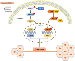
The present study was designed to explore the function of FAM172A in liver regeneration and HCC. Mice were sacrificed after 70% partial hepatectomy (PH). RNA sequencing was performed on primary hepatocytes of WT and FAM172A-/- mice. We used HepG2 cells to construct cell lines with stably knockdown and overexpression of FAM172A. The expression of FAM172A in liver tissues was investigated by immunohistochemical staining, and we also used public database to perform survival analysis and prognostic model in HCC. Compared with WT mice after PH, normalized liver weight/body weight (LW/BW) ratio and the proliferating cell nuclear antigen (PCNA) protein level of FAM172A-/- mice elevated. The DEGs were mainly enriched in inflammatory response, tumor necrosis factor production, and wound healing. FAM172A knockdown enhanced the NFκB-TNFα and pERK-YAP1-Cyclin D1 axis. FAM172A peptide inhibited proliferation of primary hepatocytes. Moreover, the low expression of FAM172A in human HCC tissues implies a lower likelihood of survival and a valid diagnostic marker for HCC. Loss of FAM172A gene promotes cell proliferation by pERK-YAP1-Cyclin D1 and pNFκB-TNFα pathways during liver regeneration after PH. FAM172A may be a favorable diagnosis marker of HCC.
期刊介绍:
Molecular and Cellular Biochemistry: An International Journal for Chemical Biology in Health and Disease publishes original research papers and short communications in all areas of the biochemical sciences, emphasizing novel findings relevant to the biochemical basis of cellular function and disease processes, as well as the mechanics of action of hormones and chemical agents. Coverage includes membrane transport, receptor mechanism, immune response, secretory processes, and cytoskeletal function, as well as biochemical structure-function relationships in the cell.
In addition to the reports of original research, the journal publishes state of the art reviews. Specific subjects covered by Molecular and Cellular Biochemistry include cellular metabolism, cellular pathophysiology, enzymology, ion transport, lipid biochemistry, membrane biochemistry, molecular biology, nuclear structure and function, and protein chemistry.


 分享
分享
 求助内容:
求助内容: 应助结果提醒方式:
 扫码关注我们
