{"title":"半月板血管化:半月板弓周围的描述性研究。","authors":"Cyrille Decante, Céline Salaud, Stéphane Lagier, Yvan Blin, Stéphane Ploteau, Antoine Hamel","doi":"10.1007/s00276-024-03400-2","DOIUrl":null,"url":null,"abstract":"<p><strong>Purpose: </strong>The meniscal vascularization remains poorly documented, particularly its origin. The aim of this cadaveric study was to describe the origin of the arterial vascularization of the menisci.</p><p><strong>Methods: </strong>This is an anatomical study on human specimens. Twenty knees were used. The average age of the subjects was 82.7 years old (56-97). Ten knees were injected with latex-neoprene and ten knees were injected with colored gelatin mixed with India ink. The same protocol for dissection was used in all cases.</p><p><strong>Results: </strong>The meniscal vascularization is provided by the genicular arteries of the knee originating from the popliteal artery. The superior medial, superior lateral, inferior medial, inferior lateral, and middle genicular arteries had constant pathways. A second middle genicular artery was found in 55% of cases. The inferior lateral genicular artery ran alongside the meniscal's periphery. The inferior medial genicular artery followed the proximal tibial metaphysis. In all dissections, a previously undocumented small artery originated from under the middle genicular arteries. This artery remained extracapsular and followed the medial meniscal periphery. This artery has been named the \"medial capsulo-meniscal artery\". The genicular arteries formed an extensive peri-articular anastomotic vascularization for the menisci and thus referred to the \"peri-meniscal arterial archs\". The lateral peri-meniscal arch was predominantly supplied by the inferior lateral genicular artery, while the medial peri-meniscal arch was mainly supplied by the medial capsulo-meniscal artery.</p><p><strong>Conclusion: </strong>The peri-meniscal arterial archs are a vascular complex formed by the genicular arteries of the knee and an artery not previously described: the \"capsulo-meniscal artery\". These archs have a constant presence but their formation and distribution is different between the medial and lateral menisci.</p>","PeriodicalId":49461,"journal":{"name":"Surgical and Radiologic Anatomy","volume":" ","pages":"1401-1409"},"PeriodicalIF":1.2000,"publicationDate":"2024-09-01","publicationTypes":"Journal Article","fieldsOfStudy":null,"isOpenAccess":false,"openAccessPdf":"","citationCount":"0","resultStr":"{\"title\":\"Vascularization of the menisci: a descriptive study of the peri-meniscal archs.\",\"authors\":\"Cyrille Decante, Céline Salaud, Stéphane Lagier, Yvan Blin, Stéphane Ploteau, Antoine Hamel\",\"doi\":\"10.1007/s00276-024-03400-2\",\"DOIUrl\":null,\"url\":null,\"abstract\":\"<p><strong>Purpose: </strong>The meniscal vascularization remains poorly documented, particularly its origin. The aim of this cadaveric study was to describe the origin of the arterial vascularization of the menisci.</p><p><strong>Methods: </strong>This is an anatomical study on human specimens. Twenty knees were used. The average age of the subjects was 82.7 years old (56-97). Ten knees were injected with latex-neoprene and ten knees were injected with colored gelatin mixed with India ink. The same protocol for dissection was used in all cases.</p><p><strong>Results: </strong>The meniscal vascularization is provided by the genicular arteries of the knee originating from the popliteal artery. The superior medial, superior lateral, inferior medial, inferior lateral, and middle genicular arteries had constant pathways. A second middle genicular artery was found in 55% of cases. The inferior lateral genicular artery ran alongside the meniscal's periphery. The inferior medial genicular artery followed the proximal tibial metaphysis. In all dissections, a previously undocumented small artery originated from under the middle genicular arteries. This artery remained extracapsular and followed the medial meniscal periphery. This artery has been named the \\\"medial capsulo-meniscal artery\\\". The genicular arteries formed an extensive peri-articular anastomotic vascularization for the menisci and thus referred to the \\\"peri-meniscal arterial archs\\\". The lateral peri-meniscal arch was predominantly supplied by the inferior lateral genicular artery, while the medial peri-meniscal arch was mainly supplied by the medial capsulo-meniscal artery.</p><p><strong>Conclusion: </strong>The peri-meniscal arterial archs are a vascular complex formed by the genicular arteries of the knee and an artery not previously described: the \\\"capsulo-meniscal artery\\\". These archs have a constant presence but their formation and distribution is different between the medial and lateral menisci.</p>\",\"PeriodicalId\":49461,\"journal\":{\"name\":\"Surgical and Radiologic Anatomy\",\"volume\":\" \",\"pages\":\"1401-1409\"},\"PeriodicalIF\":1.2000,\"publicationDate\":\"2024-09-01\",\"publicationTypes\":\"Journal Article\",\"fieldsOfStudy\":null,\"isOpenAccess\":false,\"openAccessPdf\":\"\",\"citationCount\":\"0\",\"resultStr\":null,\"platform\":\"Semanticscholar\",\"paperid\":null,\"PeriodicalName\":\"Surgical and Radiologic Anatomy\",\"FirstCategoryId\":\"3\",\"ListUrlMain\":\"https://doi.org/10.1007/s00276-024-03400-2\",\"RegionNum\":4,\"RegionCategory\":\"医学\",\"ArticlePicture\":[],\"TitleCN\":null,\"AbstractTextCN\":null,\"PMCID\":null,\"EPubDate\":\"2024/6/18 0:00:00\",\"PubModel\":\"Epub\",\"JCR\":\"Q2\",\"JCRName\":\"Medicine\",\"Score\":null,\"Total\":0}","platform":"Semanticscholar","paperid":null,"PeriodicalName":"Surgical and Radiologic Anatomy","FirstCategoryId":"3","ListUrlMain":"https://doi.org/10.1007/s00276-024-03400-2","RegionNum":4,"RegionCategory":"医学","ArticlePicture":[],"TitleCN":null,"AbstractTextCN":null,"PMCID":null,"EPubDate":"2024/6/18 0:00:00","PubModel":"Epub","JCR":"Q2","JCRName":"Medicine","Score":null,"Total":0}
Vascularization of the menisci: a descriptive study of the peri-meniscal archs.
Purpose: The meniscal vascularization remains poorly documented, particularly its origin. The aim of this cadaveric study was to describe the origin of the arterial vascularization of the menisci.
Methods: This is an anatomical study on human specimens. Twenty knees were used. The average age of the subjects was 82.7 years old (56-97). Ten knees were injected with latex-neoprene and ten knees were injected with colored gelatin mixed with India ink. The same protocol for dissection was used in all cases.
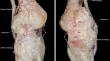
Results: The meniscal vascularization is provided by the genicular arteries of the knee originating from the popliteal artery. The superior medial, superior lateral, inferior medial, inferior lateral, and middle genicular arteries had constant pathways. A second middle genicular artery was found in 55% of cases. The inferior lateral genicular artery ran alongside the meniscal's periphery. The inferior medial genicular artery followed the proximal tibial metaphysis. In all dissections, a previously undocumented small artery originated from under the middle genicular arteries. This artery remained extracapsular and followed the medial meniscal periphery. This artery has been named the "medial capsulo-meniscal artery". The genicular arteries formed an extensive peri-articular anastomotic vascularization for the menisci and thus referred to the "peri-meniscal arterial archs". The lateral peri-meniscal arch was predominantly supplied by the inferior lateral genicular artery, while the medial peri-meniscal arch was mainly supplied by the medial capsulo-meniscal artery.
Conclusion: The peri-meniscal arterial archs are a vascular complex formed by the genicular arteries of the knee and an artery not previously described: the "capsulo-meniscal artery". These archs have a constant presence but their formation and distribution is different between the medial and lateral menisci.
期刊介绍:
Anatomy is a morphological science which cannot fail to interest the clinician. The practical application of anatomical research to clinical problems necessitates special adaptation and selectivity in choosing from numerous international works. Although there is a tendency to believe that meaningful advances in anatomy are unlikely, constant revision is necessary. Surgical and Radiologic Anatomy, the first international journal of Clinical anatomy has been created in this spirit.
Its goal is to serve clinicians, regardless of speciality-physicians, surgeons, radiologists or other specialists-as an indispensable aid with which they can improve their knowledge of anatomy. Each issue includes: Original papers, review articles, articles on the anatomical bases of medical, surgical and radiological techniques, articles of normal radiologic anatomy, brief reviews of anatomical publications of clinical interest.
Particular attention is given to high quality illustrations, which are indispensable for a better understanding of anatomical problems.
Surgical and Radiologic Anatomy is a journal written by anatomists for clinicians with a special interest in anatomy.


 分享
分享
 求助内容:
求助内容: 应助结果提醒方式:
 扫码关注我们
