{"title":"原发性肝胆粘液表皮样癌:病例报告和文献综述。","authors":"Zihan Li, Hiep Nguyen Canh, Khuyen Nguyen Thi, Kenta Takahashi, Quynh Nguyen Thi, Dong Le Thanh, Rui Yang, Yasunori Sato, Kenichi Harada","doi":"10.1007/s00795-024-00390-3","DOIUrl":null,"url":null,"abstract":"<p><p>Hepatobiliary mucoepidermoid carcinoma is a rare malignant tumor comprising mucous, intermediate, and epidermoid cells. Herein, we presented a case of primary liver mucoepidermoid carcinoma preoperatively misdiagnosed as conventional intrahepatic cholangiocarcinoma. A 67-year-old male was admitted to our hospital. Preoperative laboratory tests showed increased aspartate transaminase, alanine transaminase, and carbohydrate antigen 19-9. Abdominal Computer Tomography revealed a 4.8 × 4.9 cm liver mass in segment VI. A preliminary diagnosis of intrahepatic cholangiocarcinoma was made, with undergoing partial hepatectomy. However, on histopathology, the tumor comprised a mixture of epidermoid, mucous, and intermediate cells with diffuse infiltrating at the tumor margin. On special stains, mucous and intermedia cells were positive for mucicarmine and Alcian blue, whereas epidermoid cells were positive for Keratin 5/6 and p63. Intermediate cells are also positive for p63. All tumor cells were positive for Keratin 7. The Ki-67 index was 35%. The final diagnosis was primary hepatic mucoepidermoid carcinoma. Although rare, hepatic mucoepidermoid carcinoma should be considered in the intrahepatic cholangiocarcinoma differential diagnosis. We reviewed previous studies and found that hepatobiliary mucoepidermoid carcinoma is more likely to originate from the biliary tract adjacent to the tumor.</p>","PeriodicalId":18338,"journal":{"name":"Medical Molecular Morphology","volume":" ","pages":"233-243"},"PeriodicalIF":1.1000,"publicationDate":"2024-09-01","publicationTypes":"Journal Article","fieldsOfStudy":null,"isOpenAccess":false,"openAccessPdf":"","citationCount":"0","resultStr":"{\"title\":\"Primary hepatobiliary mucoepidermoid carcinoma: a case report and review of literature.\",\"authors\":\"Zihan Li, Hiep Nguyen Canh, Khuyen Nguyen Thi, Kenta Takahashi, Quynh Nguyen Thi, Dong Le Thanh, Rui Yang, Yasunori Sato, Kenichi Harada\",\"doi\":\"10.1007/s00795-024-00390-3\",\"DOIUrl\":null,\"url\":null,\"abstract\":\"<p><p>Hepatobiliary mucoepidermoid carcinoma is a rare malignant tumor comprising mucous, intermediate, and epidermoid cells. Herein, we presented a case of primary liver mucoepidermoid carcinoma preoperatively misdiagnosed as conventional intrahepatic cholangiocarcinoma. A 67-year-old male was admitted to our hospital. Preoperative laboratory tests showed increased aspartate transaminase, alanine transaminase, and carbohydrate antigen 19-9. Abdominal Computer Tomography revealed a 4.8 × 4.9 cm liver mass in segment VI. A preliminary diagnosis of intrahepatic cholangiocarcinoma was made, with undergoing partial hepatectomy. However, on histopathology, the tumor comprised a mixture of epidermoid, mucous, and intermediate cells with diffuse infiltrating at the tumor margin. On special stains, mucous and intermedia cells were positive for mucicarmine and Alcian blue, whereas epidermoid cells were positive for Keratin 5/6 and p63. Intermediate cells are also positive for p63. All tumor cells were positive for Keratin 7. The Ki-67 index was 35%. The final diagnosis was primary hepatic mucoepidermoid carcinoma. Although rare, hepatic mucoepidermoid carcinoma should be considered in the intrahepatic cholangiocarcinoma differential diagnosis. We reviewed previous studies and found that hepatobiliary mucoepidermoid carcinoma is more likely to originate from the biliary tract adjacent to the tumor.</p>\",\"PeriodicalId\":18338,\"journal\":{\"name\":\"Medical Molecular Morphology\",\"volume\":\" \",\"pages\":\"233-243\"},\"PeriodicalIF\":1.1000,\"publicationDate\":\"2024-09-01\",\"publicationTypes\":\"Journal Article\",\"fieldsOfStudy\":null,\"isOpenAccess\":false,\"openAccessPdf\":\"\",\"citationCount\":\"0\",\"resultStr\":null,\"platform\":\"Semanticscholar\",\"paperid\":null,\"PeriodicalName\":\"Medical Molecular Morphology\",\"FirstCategoryId\":\"3\",\"ListUrlMain\":\"https://doi.org/10.1007/s00795-024-00390-3\",\"RegionNum\":4,\"RegionCategory\":\"医学\",\"ArticlePicture\":[],\"TitleCN\":null,\"AbstractTextCN\":null,\"PMCID\":null,\"EPubDate\":\"2024/6/21 0:00:00\",\"PubModel\":\"Epub\",\"JCR\":\"Q3\",\"JCRName\":\"PATHOLOGY\",\"Score\":null,\"Total\":0}","platform":"Semanticscholar","paperid":null,"PeriodicalName":"Medical Molecular Morphology","FirstCategoryId":"3","ListUrlMain":"https://doi.org/10.1007/s00795-024-00390-3","RegionNum":4,"RegionCategory":"医学","ArticlePicture":[],"TitleCN":null,"AbstractTextCN":null,"PMCID":null,"EPubDate":"2024/6/21 0:00:00","PubModel":"Epub","JCR":"Q3","JCRName":"PATHOLOGY","Score":null,"Total":0}
Primary hepatobiliary mucoepidermoid carcinoma: a case report and review of literature.
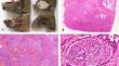
Hepatobiliary mucoepidermoid carcinoma is a rare malignant tumor comprising mucous, intermediate, and epidermoid cells. Herein, we presented a case of primary liver mucoepidermoid carcinoma preoperatively misdiagnosed as conventional intrahepatic cholangiocarcinoma. A 67-year-old male was admitted to our hospital. Preoperative laboratory tests showed increased aspartate transaminase, alanine transaminase, and carbohydrate antigen 19-9. Abdominal Computer Tomography revealed a 4.8 × 4.9 cm liver mass in segment VI. A preliminary diagnosis of intrahepatic cholangiocarcinoma was made, with undergoing partial hepatectomy. However, on histopathology, the tumor comprised a mixture of epidermoid, mucous, and intermediate cells with diffuse infiltrating at the tumor margin. On special stains, mucous and intermedia cells were positive for mucicarmine and Alcian blue, whereas epidermoid cells were positive for Keratin 5/6 and p63. Intermediate cells are also positive for p63. All tumor cells were positive for Keratin 7. The Ki-67 index was 35%. The final diagnosis was primary hepatic mucoepidermoid carcinoma. Although rare, hepatic mucoepidermoid carcinoma should be considered in the intrahepatic cholangiocarcinoma differential diagnosis. We reviewed previous studies and found that hepatobiliary mucoepidermoid carcinoma is more likely to originate from the biliary tract adjacent to the tumor.
期刊介绍:
Medical Molecular Morphology is an international forum for researchers in both basic and clinical medicine to present and discuss new research on the structural mechanisms and the processes of health and disease at the molecular level. The structures of molecules, organelles, cells, tissues, and organs determine their normal function. Disease is thus best understood in terms of structural changes in these different levels of biological organization, especially in molecules and molecular interactions as well as the cellular localization of chemical components. Medical Molecular Morphology welcomes articles on basic or clinical research in the fields of cell biology, molecular biology, and medical, veterinary, and dental sciences using techniques for structural research such as electron microscopy, confocal laser scanning microscopy, enzyme histochemistry, immunohistochemistry, radioautography, X-ray microanalysis, and in situ hybridization.
Manuscripts submitted for publication must contain a statement to the effect that all human studies have been reviewed by the appropriate ethics committee and have therefore been performed in accordance with the ethical standards laid down in an appropriate version of the 1964 Declaration of Helsinki. It should also be stated clearly in the text that all persons gave their informed consent prior to their inclusion in the study. Details that might disclose the identity of the subjects under study should be omitted.


 分享
分享
 求助内容:
求助内容: 应助结果提醒方式:
 扫码关注我们
