Simin Liu, Yao Zhang, Wei Liu, Ting Yin, Jie Yuan, Jun Ran, Xiaoming Li
{"title":"缩短膝关节弥散张量成像采集时间的同步多切片技术:一项可行性研究。","authors":"Simin Liu, Yao Zhang, Wei Liu, Ting Yin, Jie Yuan, Jun Ran, Xiaoming Li","doi":"10.1007/s00256-024-04719-y","DOIUrl":null,"url":null,"abstract":"<p><strong>Objectives: </strong>To explore the feasibility of simultaneous multi-slice (SMS) technique for reducing acquisition times in readout-segmented echo planar imaging (RESOLVE) for diffusion tensor imaging (DTI) of the knee.</p><p><strong>Materials and methods: </strong>A total of 30 healthy volunteers and 23 patients with knee acute injury (12 cases with anterior ligament (ACL) tears and 16 cases with patellar cartilage (PC) injury) were enrolled in this prospective study. Three DTI protocols were used: conventional RESOLVE-DTI with 12 directions (protocol 1), SMS-RESOLVE-DTI with 12 directions (protocol 2) and 20 directions (protocol 3). DTI parameters of gastrocnemius, ACL and posterior cruciate ligament (PCL), and PC from three protocols were quantitatively assessed.</p><p><strong>Results: </strong>For volunteers, protocol 2 significantly reduced acquisition time by 38.6% and 34.2% compared to protocols 1 and 3 while maintaining similar high-quality images and similar diffusive parameters, except for the fractional anisotropy (FA) and axial diffusivity (AD) of the PC between protocols 2 and 1 (P < 0.05). For injured ACL and PC, protocols 1 and 2 showed similar accurate diffusive parameters (except for AD, P = 0.025) and similar diagnostic efficacy, which demonstrated significantly lower FA and higher radial diffusivity (RD) in protocols 1 and 2 compared to volunteers (P < 0.05).</p><p><strong>Conclusions: </strong>The 12-direction SMS-RESOLVE-DTI demonstrated a favorable balance between acquisition time and image quality, making it a promising alternative to conventional DTI for evaluating ligament and cartilage injuries.</p><p><strong>Advances in knowledge: </strong>The SMS technique greatly reduces acquisition time while maintaining image quality, which signified the possibility of DTI's clinical application.</p>","PeriodicalId":21783,"journal":{"name":"Skeletal Radiology","volume":" ","pages":"243-253"},"PeriodicalIF":2.2000,"publicationDate":"2025-02-01","publicationTypes":"Journal Article","fieldsOfStudy":null,"isOpenAccess":false,"openAccessPdf":"","citationCount":"0","resultStr":"{\"title\":\"Simultaneous multi-slice technique for reducing acquisition times in diffusion tensor imaging of the knee: a feasibility study.\",\"authors\":\"Simin Liu, Yao Zhang, Wei Liu, Ting Yin, Jie Yuan, Jun Ran, Xiaoming Li\",\"doi\":\"10.1007/s00256-024-04719-y\",\"DOIUrl\":null,\"url\":null,\"abstract\":\"<p><strong>Objectives: </strong>To explore the feasibility of simultaneous multi-slice (SMS) technique for reducing acquisition times in readout-segmented echo planar imaging (RESOLVE) for diffusion tensor imaging (DTI) of the knee.</p><p><strong>Materials and methods: </strong>A total of 30 healthy volunteers and 23 patients with knee acute injury (12 cases with anterior ligament (ACL) tears and 16 cases with patellar cartilage (PC) injury) were enrolled in this prospective study. Three DTI protocols were used: conventional RESOLVE-DTI with 12 directions (protocol 1), SMS-RESOLVE-DTI with 12 directions (protocol 2) and 20 directions (protocol 3). DTI parameters of gastrocnemius, ACL and posterior cruciate ligament (PCL), and PC from three protocols were quantitatively assessed.</p><p><strong>Results: </strong>For volunteers, protocol 2 significantly reduced acquisition time by 38.6% and 34.2% compared to protocols 1 and 3 while maintaining similar high-quality images and similar diffusive parameters, except for the fractional anisotropy (FA) and axial diffusivity (AD) of the PC between protocols 2 and 1 (P < 0.05). For injured ACL and PC, protocols 1 and 2 showed similar accurate diffusive parameters (except for AD, P = 0.025) and similar diagnostic efficacy, which demonstrated significantly lower FA and higher radial diffusivity (RD) in protocols 1 and 2 compared to volunteers (P < 0.05).</p><p><strong>Conclusions: </strong>The 12-direction SMS-RESOLVE-DTI demonstrated a favorable balance between acquisition time and image quality, making it a promising alternative to conventional DTI for evaluating ligament and cartilage injuries.</p><p><strong>Advances in knowledge: </strong>The SMS technique greatly reduces acquisition time while maintaining image quality, which signified the possibility of DTI's clinical application.</p>\",\"PeriodicalId\":21783,\"journal\":{\"name\":\"Skeletal Radiology\",\"volume\":\" \",\"pages\":\"243-253\"},\"PeriodicalIF\":2.2000,\"publicationDate\":\"2025-02-01\",\"publicationTypes\":\"Journal Article\",\"fieldsOfStudy\":null,\"isOpenAccess\":false,\"openAccessPdf\":\"\",\"citationCount\":\"0\",\"resultStr\":null,\"platform\":\"Semanticscholar\",\"paperid\":null,\"PeriodicalName\":\"Skeletal Radiology\",\"FirstCategoryId\":\"3\",\"ListUrlMain\":\"https://doi.org/10.1007/s00256-024-04719-y\",\"RegionNum\":3,\"RegionCategory\":\"医学\",\"ArticlePicture\":[],\"TitleCN\":null,\"AbstractTextCN\":null,\"PMCID\":null,\"EPubDate\":\"2024/6/24 0:00:00\",\"PubModel\":\"Epub\",\"JCR\":\"Q2\",\"JCRName\":\"ORTHOPEDICS\",\"Score\":null,\"Total\":0}","platform":"Semanticscholar","paperid":null,"PeriodicalName":"Skeletal Radiology","FirstCategoryId":"3","ListUrlMain":"https://doi.org/10.1007/s00256-024-04719-y","RegionNum":3,"RegionCategory":"医学","ArticlePicture":[],"TitleCN":null,"AbstractTextCN":null,"PMCID":null,"EPubDate":"2024/6/24 0:00:00","PubModel":"Epub","JCR":"Q2","JCRName":"ORTHOPEDICS","Score":null,"Total":0}
Simultaneous multi-slice technique for reducing acquisition times in diffusion tensor imaging of the knee: a feasibility study.
Objectives: To explore the feasibility of simultaneous multi-slice (SMS) technique for reducing acquisition times in readout-segmented echo planar imaging (RESOLVE) for diffusion tensor imaging (DTI) of the knee.
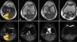
Materials and methods: A total of 30 healthy volunteers and 23 patients with knee acute injury (12 cases with anterior ligament (ACL) tears and 16 cases with patellar cartilage (PC) injury) were enrolled in this prospective study. Three DTI protocols were used: conventional RESOLVE-DTI with 12 directions (protocol 1), SMS-RESOLVE-DTI with 12 directions (protocol 2) and 20 directions (protocol 3). DTI parameters of gastrocnemius, ACL and posterior cruciate ligament (PCL), and PC from three protocols were quantitatively assessed.
Results: For volunteers, protocol 2 significantly reduced acquisition time by 38.6% and 34.2% compared to protocols 1 and 3 while maintaining similar high-quality images and similar diffusive parameters, except for the fractional anisotropy (FA) and axial diffusivity (AD) of the PC between protocols 2 and 1 (P < 0.05). For injured ACL and PC, protocols 1 and 2 showed similar accurate diffusive parameters (except for AD, P = 0.025) and similar diagnostic efficacy, which demonstrated significantly lower FA and higher radial diffusivity (RD) in protocols 1 and 2 compared to volunteers (P < 0.05).
Conclusions: The 12-direction SMS-RESOLVE-DTI demonstrated a favorable balance between acquisition time and image quality, making it a promising alternative to conventional DTI for evaluating ligament and cartilage injuries.
Advances in knowledge: The SMS technique greatly reduces acquisition time while maintaining image quality, which signified the possibility of DTI's clinical application.
期刊介绍:
Skeletal Radiology provides a forum for the dissemination of current knowledge and information dealing with disorders of the musculoskeletal system including the spine. While emphasizing the radiological aspects of the many varied skeletal abnormalities, the journal also adopts an interdisciplinary approach, reflecting the membership of the International Skeletal Society. Thus, the anatomical, pathological, physiological, clinical, metabolic and epidemiological aspects of the many entities affecting the skeleton receive appropriate consideration.
This is the Journal of the International Skeletal Society and the Official Journal of the Society of Skeletal Radiology and the Australasian Musculoskelelal Imaging Group.


 分享
分享
 求助内容:
求助内容: 应助结果提醒方式:
 扫码关注我们
