Mingming Cai, Wangdu Luo, Kevin Feng, Yi Chen, Lin Yi, Xiaomin Zhu, Ju He, Hong Liu, Cindy Hutnik, Yong Wang, Xiangji Li, Lin Xie
{"title":"将 HA-Mg 青光眼引流板植入兔眼前房的眼压控制效果和安全性。","authors":"Mingming Cai, Wangdu Luo, Kevin Feng, Yi Chen, Lin Yi, Xiaomin Zhu, Ju He, Hong Liu, Cindy Hutnik, Yong Wang, Xiangji Li, Lin Xie","doi":"10.1007/s10856-024-06806-x","DOIUrl":null,"url":null,"abstract":"<p>The current clinical application of glaucoma drainage devices is made of non-degradable materials. These non-degradable drainage devices often trigger inflammatory responses and scar proliferation, possibly leading to surgical failure. We developed a biodegradable material hydroxyapatite-coated magnesium (HA-Mg) as a glaucoma drainage device. Twelve New Zealand white rabbits were randomly assigned to three groups: HA-Mg drainage plate group (6 right eyes), trabeculectomy group (6 right eyes), and control group (12 left eyes). Results showed that all HA-Mg drainage plates were completely degraded ~4 months postoperatively. At the 5th month postoperatively, there was no statistical difference in the corneal endothelium density between the HA-Mg drainage plate group and the control group (<i>p</i> = 0.857). The intraocular pressure (IOP) level in the HA-Mg drainage plate implantation group was lower than in the other two groups. The trypan blue dye still drained from the anterior chamber to the subconjunctiva 5 months after HA-Mg drainage plate implantation. HE staining revealed the scleral linear aqueous humor drainage channel and anterior synechia were observed after drainage plate completely degraded, with no obvious infiltration with the inflammatory cells. This study showed the safety and efficacy of HA-Mg glaucoma drainage plate in controlling IOP after implantation into the anterior chamber of rabbit eyes.</p>","PeriodicalId":647,"journal":{"name":"Journal of Materials Science: Materials in Medicine","volume":"35 1","pages":""},"PeriodicalIF":4.5000,"publicationDate":"2024-06-25","publicationTypes":"Journal Article","fieldsOfStudy":null,"isOpenAccess":false,"openAccessPdf":"https://www.ncbi.nlm.nih.gov/pmc/articles/PMC11199312/pdf/","citationCount":"0","resultStr":"{\"title\":\"Intraocular pressure control efficacy and safety of HA-Mg glaucoma drainage plate implantation in the anterior chamber of rabbit eyes\",\"authors\":\"Mingming Cai, Wangdu Luo, Kevin Feng, Yi Chen, Lin Yi, Xiaomin Zhu, Ju He, Hong Liu, Cindy Hutnik, Yong Wang, Xiangji Li, Lin Xie\",\"doi\":\"10.1007/s10856-024-06806-x\",\"DOIUrl\":null,\"url\":null,\"abstract\":\"<p>The current clinical application of glaucoma drainage devices is made of non-degradable materials. These non-degradable drainage devices often trigger inflammatory responses and scar proliferation, possibly leading to surgical failure. We developed a biodegradable material hydroxyapatite-coated magnesium (HA-Mg) as a glaucoma drainage device. Twelve New Zealand white rabbits were randomly assigned to three groups: HA-Mg drainage plate group (6 right eyes), trabeculectomy group (6 right eyes), and control group (12 left eyes). Results showed that all HA-Mg drainage plates were completely degraded ~4 months postoperatively. At the 5th month postoperatively, there was no statistical difference in the corneal endothelium density between the HA-Mg drainage plate group and the control group (<i>p</i> = 0.857). The intraocular pressure (IOP) level in the HA-Mg drainage plate implantation group was lower than in the other two groups. The trypan blue dye still drained from the anterior chamber to the subconjunctiva 5 months after HA-Mg drainage plate implantation. HE staining revealed the scleral linear aqueous humor drainage channel and anterior synechia were observed after drainage plate completely degraded, with no obvious infiltration with the inflammatory cells. This study showed the safety and efficacy of HA-Mg glaucoma drainage plate in controlling IOP after implantation into the anterior chamber of rabbit eyes.</p>\",\"PeriodicalId\":647,\"journal\":{\"name\":\"Journal of Materials Science: Materials in Medicine\",\"volume\":\"35 1\",\"pages\":\"\"},\"PeriodicalIF\":4.5000,\"publicationDate\":\"2024-06-25\",\"publicationTypes\":\"Journal Article\",\"fieldsOfStudy\":null,\"isOpenAccess\":false,\"openAccessPdf\":\"https://www.ncbi.nlm.nih.gov/pmc/articles/PMC11199312/pdf/\",\"citationCount\":\"0\",\"resultStr\":null,\"platform\":\"Semanticscholar\",\"paperid\":null,\"PeriodicalName\":\"Journal of Materials Science: Materials in Medicine\",\"FirstCategoryId\":\"5\",\"ListUrlMain\":\"https://link.springer.com/article/10.1007/s10856-024-06806-x\",\"RegionNum\":3,\"RegionCategory\":\"医学\",\"ArticlePicture\":[],\"TitleCN\":null,\"AbstractTextCN\":null,\"PMCID\":null,\"EPubDate\":\"\",\"PubModel\":\"\",\"JCR\":\"Q2\",\"JCRName\":\"ENGINEERING, BIOMEDICAL\",\"Score\":null,\"Total\":0}","platform":"Semanticscholar","paperid":null,"PeriodicalName":"Journal of Materials Science: Materials in Medicine","FirstCategoryId":"5","ListUrlMain":"https://link.springer.com/article/10.1007/s10856-024-06806-x","RegionNum":3,"RegionCategory":"医学","ArticlePicture":[],"TitleCN":null,"AbstractTextCN":null,"PMCID":null,"EPubDate":"","PubModel":"","JCR":"Q2","JCRName":"ENGINEERING, BIOMEDICAL","Score":null,"Total":0}
Intraocular pressure control efficacy and safety of HA-Mg glaucoma drainage plate implantation in the anterior chamber of rabbit eyes
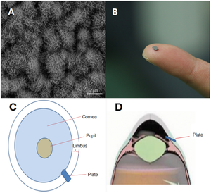
The current clinical application of glaucoma drainage devices is made of non-degradable materials. These non-degradable drainage devices often trigger inflammatory responses and scar proliferation, possibly leading to surgical failure. We developed a biodegradable material hydroxyapatite-coated magnesium (HA-Mg) as a glaucoma drainage device. Twelve New Zealand white rabbits were randomly assigned to three groups: HA-Mg drainage plate group (6 right eyes), trabeculectomy group (6 right eyes), and control group (12 left eyes). Results showed that all HA-Mg drainage plates were completely degraded ~4 months postoperatively. At the 5th month postoperatively, there was no statistical difference in the corneal endothelium density between the HA-Mg drainage plate group and the control group (p = 0.857). The intraocular pressure (IOP) level in the HA-Mg drainage plate implantation group was lower than in the other two groups. The trypan blue dye still drained from the anterior chamber to the subconjunctiva 5 months after HA-Mg drainage plate implantation. HE staining revealed the scleral linear aqueous humor drainage channel and anterior synechia were observed after drainage plate completely degraded, with no obvious infiltration with the inflammatory cells. This study showed the safety and efficacy of HA-Mg glaucoma drainage plate in controlling IOP after implantation into the anterior chamber of rabbit eyes.
期刊介绍:
The Journal of Materials Science: Materials in Medicine publishes refereed papers providing significant progress in the application of biomaterials and tissue engineering constructs as medical or dental implants, prostheses and devices. Coverage spans a wide range of topics from basic science to clinical applications, around the theme of materials in medicine and dentistry. The central element is the development of synthetic and natural materials used in orthopaedic, maxillofacial, cardiovascular, neurological, ophthalmic and dental applications. Special biomedical topics include biomaterial synthesis and characterisation, biocompatibility studies, nanomedicine, tissue engineering constructs and cell substrates, regenerative medicine, computer modelling and other advanced experimental methodologies.


 分享
分享
 求助内容:
求助内容: 应助结果提醒方式:
 扫码关注我们
