Kai Cheng, Haotian Zhu, Yuanhao Peng, Xinghua Wen, Huanwen Ding
{"title":"计算机辅助设计和三维打印技术用于稳定构建比格犬节段骨缺损模型:短期观察。","authors":"Kai Cheng, Haotian Zhu, Yuanhao Peng, Xinghua Wen, Huanwen Ding","doi":"10.1186/s41205-024-00217-y","DOIUrl":null,"url":null,"abstract":"<p><strong>Objective: </strong>Segmental bone defect animal studies require stable fixation which is a continuous experimental challenge. Large animal models are comparable to the human bone, but with obvious drawbacks of housing and costs. Our study aims to utilize CAD and 3D printing in the construction of a stable and reproducible segmental bone defect animal mode.</p><p><strong>Methods: </strong>CAD-aided 3D printed surgical instruments were incorporated into the construction of the animal model through preoperative surgical emulation. 20 3D printed femurs were divided into either experimental group using 3D surgical instruments or control group. In Vitro surgical time and accuracy of fixation were analysed and compared between the two groups. A mature surgical plan using the surgical instruments was then utilized in the construction of 3 segmental bone defect Beagle models in vivo. The Beagles were postoperatively assessed through limb function and imaging at 1, 2 and 3 months postoperatively.</p><p><strong>Results: </strong>In vitro experiments showed a significant reduction in surgical time from 40.6 ± 14.1 (23-68 min) to 26 ± 4.6 (19-36 min) (n = 10, p < 0.05) and the accuracy of intramedullary fixation placement increased from 71.6 ± 23.6 (33.3-100) % to 98.3 ± 5.37 (83-100) %, (n = 30, p < 0.05) with the use of CAD and 3D printed instruments. All Beagles were load-bearing within 1 week, and postoperative radiographs showed no evidence of implant failure.</p><p><strong>Conclusion: </strong>Incorporation of CAD and 3D printing significantly increases stability, while reducing the surgical time in the construction of the animal model, significantly affecting the success of the segmental bone defect model in Beagles.</p>","PeriodicalId":72036,"journal":{"name":"3D printing in medicine","volume":"10 1","pages":"20"},"PeriodicalIF":3.1000,"publicationDate":"2024-06-25","publicationTypes":"Journal Article","fieldsOfStudy":null,"isOpenAccess":false,"openAccessPdf":"https://www.ncbi.nlm.nih.gov/pmc/articles/PMC11197206/pdf/","citationCount":"0","resultStr":"{\"title\":\"Computer-aided design and 3D printing for a stable construction of segmental bone defect model in Beagles: a short term observation.\",\"authors\":\"Kai Cheng, Haotian Zhu, Yuanhao Peng, Xinghua Wen, Huanwen Ding\",\"doi\":\"10.1186/s41205-024-00217-y\",\"DOIUrl\":null,\"url\":null,\"abstract\":\"<p><strong>Objective: </strong>Segmental bone defect animal studies require stable fixation which is a continuous experimental challenge. Large animal models are comparable to the human bone, but with obvious drawbacks of housing and costs. Our study aims to utilize CAD and 3D printing in the construction of a stable and reproducible segmental bone defect animal mode.</p><p><strong>Methods: </strong>CAD-aided 3D printed surgical instruments were incorporated into the construction of the animal model through preoperative surgical emulation. 20 3D printed femurs were divided into either experimental group using 3D surgical instruments or control group. In Vitro surgical time and accuracy of fixation were analysed and compared between the two groups. A mature surgical plan using the surgical instruments was then utilized in the construction of 3 segmental bone defect Beagle models in vivo. The Beagles were postoperatively assessed through limb function and imaging at 1, 2 and 3 months postoperatively.</p><p><strong>Results: </strong>In vitro experiments showed a significant reduction in surgical time from 40.6 ± 14.1 (23-68 min) to 26 ± 4.6 (19-36 min) (n = 10, p < 0.05) and the accuracy of intramedullary fixation placement increased from 71.6 ± 23.6 (33.3-100) % to 98.3 ± 5.37 (83-100) %, (n = 30, p < 0.05) with the use of CAD and 3D printed instruments. All Beagles were load-bearing within 1 week, and postoperative radiographs showed no evidence of implant failure.</p><p><strong>Conclusion: </strong>Incorporation of CAD and 3D printing significantly increases stability, while reducing the surgical time in the construction of the animal model, significantly affecting the success of the segmental bone defect model in Beagles.</p>\",\"PeriodicalId\":72036,\"journal\":{\"name\":\"3D printing in medicine\",\"volume\":\"10 1\",\"pages\":\"20\"},\"PeriodicalIF\":3.1000,\"publicationDate\":\"2024-06-25\",\"publicationTypes\":\"Journal Article\",\"fieldsOfStudy\":null,\"isOpenAccess\":false,\"openAccessPdf\":\"https://www.ncbi.nlm.nih.gov/pmc/articles/PMC11197206/pdf/\",\"citationCount\":\"0\",\"resultStr\":null,\"platform\":\"Semanticscholar\",\"paperid\":null,\"PeriodicalName\":\"3D printing in medicine\",\"FirstCategoryId\":\"1085\",\"ListUrlMain\":\"https://doi.org/10.1186/s41205-024-00217-y\",\"RegionNum\":0,\"RegionCategory\":null,\"ArticlePicture\":[],\"TitleCN\":null,\"AbstractTextCN\":null,\"PMCID\":null,\"EPubDate\":\"\",\"PubModel\":\"\",\"JCR\":\"Q1\",\"JCRName\":\"RADIOLOGY, NUCLEAR MEDICINE & MEDICAL IMAGING\",\"Score\":null,\"Total\":0}","platform":"Semanticscholar","paperid":null,"PeriodicalName":"3D printing in medicine","FirstCategoryId":"1085","ListUrlMain":"https://doi.org/10.1186/s41205-024-00217-y","RegionNum":0,"RegionCategory":null,"ArticlePicture":[],"TitleCN":null,"AbstractTextCN":null,"PMCID":null,"EPubDate":"","PubModel":"","JCR":"Q1","JCRName":"RADIOLOGY, NUCLEAR MEDICINE & MEDICAL IMAGING","Score":null,"Total":0}
引用次数: 0
摘要
目的:节段性骨缺损动物研究需要稳定的固定,这是一项持续的实验挑战。大型动物模型可与人体骨骼媲美,但在房舍和成本方面存在明显缺陷。我们的研究旨在利用 CAD 和 3D 打印技术构建稳定、可重复的节段性骨缺损动物模型:方法:通过术前手术模拟,将 CAD 辅助的 3D 打印手术器械纳入动物模型的构建中。20 个 3D 打印股骨被分为使用 3D 手术器械的实验组和对照组。对两组的体外手术时间和固定准确性进行了分析和比较。然后,使用手术器械制定了成熟的手术方案,在体内构建了 3 个节段性骨缺损比格犬模型。术后通过肢体功能和术后 1、2 和 3 个月的成像对比格犬进行评估:结果:体外实验显示,手术时间从 40.6 ± 14.1 (23-68 分钟) 显著缩短至 26 ± 4.6 (19-36 分钟) (n = 10, p 结论:CAD 和 3D 打印技术的结合可显著缩短手术时间:在构建动物模型的过程中,CAD 和 3D 打印技术的应用大大提高了稳定性,同时缩短了手术时间,对比格犬节段骨缺损模型的成功率有显著影响。
Computer-aided design and 3D printing for a stable construction of segmental bone defect model in Beagles: a short term observation.
Objective: Segmental bone defect animal studies require stable fixation which is a continuous experimental challenge. Large animal models are comparable to the human bone, but with obvious drawbacks of housing and costs. Our study aims to utilize CAD and 3D printing in the construction of a stable and reproducible segmental bone defect animal mode.
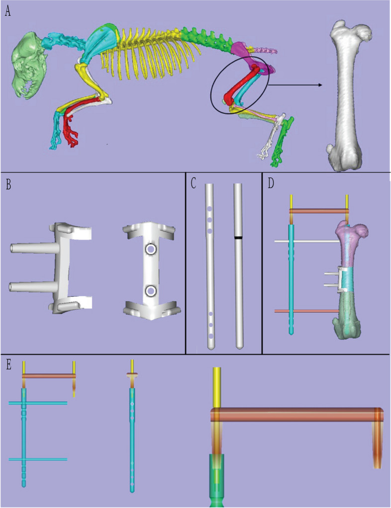
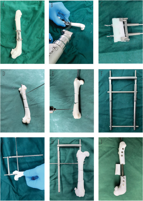
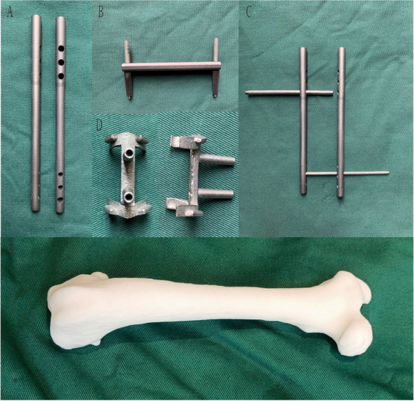
Methods: CAD-aided 3D printed surgical instruments were incorporated into the construction of the animal model through preoperative surgical emulation. 20 3D printed femurs were divided into either experimental group using 3D surgical instruments or control group. In Vitro surgical time and accuracy of fixation were analysed and compared between the two groups. A mature surgical plan using the surgical instruments was then utilized in the construction of 3 segmental bone defect Beagle models in vivo. The Beagles were postoperatively assessed through limb function and imaging at 1, 2 and 3 months postoperatively.
Results: In vitro experiments showed a significant reduction in surgical time from 40.6 ± 14.1 (23-68 min) to 26 ± 4.6 (19-36 min) (n = 10, p < 0.05) and the accuracy of intramedullary fixation placement increased from 71.6 ± 23.6 (33.3-100) % to 98.3 ± 5.37 (83-100) %, (n = 30, p < 0.05) with the use of CAD and 3D printed instruments. All Beagles were load-bearing within 1 week, and postoperative radiographs showed no evidence of implant failure.
Conclusion: Incorporation of CAD and 3D printing significantly increases stability, while reducing the surgical time in the construction of the animal model, significantly affecting the success of the segmental bone defect model in Beagles.


 分享
分享

 求助内容:
求助内容: 应助结果提醒方式:
 扫码关注我们
