Ingrid S Tam, Mohamed Elemary, John DeCoteau, Anna Porwit, Emina E Torlakovic
{"title":"COVID-19 诱导的伴有单核细胞增多的一过性红细胞白血病反应中急性单核细胞白血病的形态学线索","authors":"Ingrid S Tam, Mohamed Elemary, John DeCoteau, Anna Porwit, Emina E Torlakovic","doi":"10.3390/hematolrep16020033","DOIUrl":null,"url":null,"abstract":"<p><p>Viral infections, including those caused by COVID-19, can produce striking morphologic changes in peripheral blood. Distinguishing between reactive changes and abnormal morphology of monocytes remains particularly difficult, with low consensus rates reported amongst hematopathologists. Here, we report a patient who developed transient monocytosis of 11.06 × 10<sup>9</sup>/L with 32% promonocytes and 1% blasts during hospitalization that was secondary to severe COVID-19 infection. Three days later, the clinical status of the patient improved and the WBC had decreased to 8.47 × 10<sup>9</sup>/L with 2.2 × 10<sup>9</sup>/L monocytes. Flow cytometry studies did not reveal immunophenotypic findings specific for an overt malignant population. At no time during admission did the patient develop cytopenia(s), and she was discharged upon clinical improvement. However, the peripheral blood sample containing promonocytes was sent for molecular testing with an extended next-generation sequencing myeloid panel and was positive for pathogenic <i>NPM1</i> Type A and <i>DNMT3A</i> R882H mutations. Subsequently, despite an essentially normal complete blood count, the patient underwent a bone marrow assessment that showed acute myeloid leukemia with 77% promonocytes. This case emphasizes the critical importance of a full work up to exclude acute leukemia when classical promonocyte morphology is encountered in the peripheral blood. Promonocytes are not a part of the reactive changes associated with COVID-19 and remain specific to myeloid neoplasia.</p>","PeriodicalId":12829,"journal":{"name":"Hematology Reports","volume":"16 2","pages":"331-335"},"PeriodicalIF":1.2000,"publicationDate":"2024-05-28","publicationTypes":"Journal Article","fieldsOfStudy":null,"isOpenAccess":false,"openAccessPdf":"https://www.ncbi.nlm.nih.gov/pmc/articles/PMC11203109/pdf/","citationCount":"0","resultStr":"{\"title\":\"Morphological Clues of Acute Monocytic Leukemia in COVID-19-Induced Transient Leukoerythroblastic Reaction with Monocytosis.\",\"authors\":\"Ingrid S Tam, Mohamed Elemary, John DeCoteau, Anna Porwit, Emina E Torlakovic\",\"doi\":\"10.3390/hematolrep16020033\",\"DOIUrl\":null,\"url\":null,\"abstract\":\"<p><p>Viral infections, including those caused by COVID-19, can produce striking morphologic changes in peripheral blood. Distinguishing between reactive changes and abnormal morphology of monocytes remains particularly difficult, with low consensus rates reported amongst hematopathologists. Here, we report a patient who developed transient monocytosis of 11.06 × 10<sup>9</sup>/L with 32% promonocytes and 1% blasts during hospitalization that was secondary to severe COVID-19 infection. Three days later, the clinical status of the patient improved and the WBC had decreased to 8.47 × 10<sup>9</sup>/L with 2.2 × 10<sup>9</sup>/L monocytes. Flow cytometry studies did not reveal immunophenotypic findings specific for an overt malignant population. At no time during admission did the patient develop cytopenia(s), and she was discharged upon clinical improvement. However, the peripheral blood sample containing promonocytes was sent for molecular testing with an extended next-generation sequencing myeloid panel and was positive for pathogenic <i>NPM1</i> Type A and <i>DNMT3A</i> R882H mutations. Subsequently, despite an essentially normal complete blood count, the patient underwent a bone marrow assessment that showed acute myeloid leukemia with 77% promonocytes. This case emphasizes the critical importance of a full work up to exclude acute leukemia when classical promonocyte morphology is encountered in the peripheral blood. Promonocytes are not a part of the reactive changes associated with COVID-19 and remain specific to myeloid neoplasia.</p>\",\"PeriodicalId\":12829,\"journal\":{\"name\":\"Hematology Reports\",\"volume\":\"16 2\",\"pages\":\"331-335\"},\"PeriodicalIF\":1.2000,\"publicationDate\":\"2024-05-28\",\"publicationTypes\":\"Journal Article\",\"fieldsOfStudy\":null,\"isOpenAccess\":false,\"openAccessPdf\":\"https://www.ncbi.nlm.nih.gov/pmc/articles/PMC11203109/pdf/\",\"citationCount\":\"0\",\"resultStr\":null,\"platform\":\"Semanticscholar\",\"paperid\":null,\"PeriodicalName\":\"Hematology Reports\",\"FirstCategoryId\":\"1085\",\"ListUrlMain\":\"https://doi.org/10.3390/hematolrep16020033\",\"RegionNum\":0,\"RegionCategory\":null,\"ArticlePicture\":[],\"TitleCN\":null,\"AbstractTextCN\":null,\"PMCID\":null,\"EPubDate\":\"\",\"PubModel\":\"\",\"JCR\":\"Q4\",\"JCRName\":\"HEMATOLOGY\",\"Score\":null,\"Total\":0}","platform":"Semanticscholar","paperid":null,"PeriodicalName":"Hematology Reports","FirstCategoryId":"1085","ListUrlMain":"https://doi.org/10.3390/hematolrep16020033","RegionNum":0,"RegionCategory":null,"ArticlePicture":[],"TitleCN":null,"AbstractTextCN":null,"PMCID":null,"EPubDate":"","PubModel":"","JCR":"Q4","JCRName":"HEMATOLOGY","Score":null,"Total":0}
Morphological Clues of Acute Monocytic Leukemia in COVID-19-Induced Transient Leukoerythroblastic Reaction with Monocytosis.
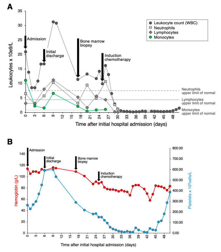
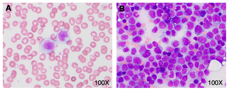
Viral infections, including those caused by COVID-19, can produce striking morphologic changes in peripheral blood. Distinguishing between reactive changes and abnormal morphology of monocytes remains particularly difficult, with low consensus rates reported amongst hematopathologists. Here, we report a patient who developed transient monocytosis of 11.06 × 10⁹/L with 32% promonocytes and 1% blasts during hospitalization that was secondary to severe COVID-19 infection. Three days later, the clinical status of the patient improved and the WBC had decreased to 8.47 × 10⁹/L with 2.2 × 10⁹/L monocytes. Flow cytometry studies did not reveal immunophenotypic findings specific for an overt malignant population. At no time during admission did the patient develop cytopenia(s), and she was discharged upon clinical improvement. However, the peripheral blood sample containing promonocytes was sent for molecular testing with an extended next-generation sequencing myeloid panel and was positive for pathogenic NPM1 Type A and DNMT3A R882H mutations. Subsequently, despite an essentially normal complete blood count, the patient underwent a bone marrow assessment that showed acute myeloid leukemia with 77% promonocytes. This case emphasizes the critical importance of a full work up to exclude acute leukemia when classical promonocyte morphology is encountered in the peripheral blood. Promonocytes are not a part of the reactive changes associated with COVID-19 and remain specific to myeloid neoplasia.


 分享
分享
 求助内容:
求助内容: 应助结果提醒方式:
 扫码关注我们
